Fully-automatic three-dimensional liver segmentation method based on local apriori information and convex optimization

A priori information, 3D liver technology, applied in the field of medical image processing, can solve the problems of under-segmentation and low accuracy of liver edge segmentation

Active Publication Date: 2016-10-26
ZHEJIANG DE IMAGE SOLUTIONS CO LTD
View PDF9 Cites 14 Cited by
  • Summary
  • Abstract
  • Description
  • Claims
  • Application Information

AI Technical Summary

Problems solved by technology

However, since the shape and position of the liver vary greatly among different individuals, the liver segmentation results obtained through the c

Method used


Image

  • Fully-automatic three-dimensional liver segmentation method based on local apriori information and convex optimization
  • Fully-automatic three-dimensional liver segmentation method based on local apriori information and convex optimization
  • Fully-automatic three-dimensional liver segmentation method based on local apriori information and convex optimization

Examples


Example Embodiment

[0049] The present invention will be further described in detail below in conjunction with the drawings and specific embodiments:

[0050] The following embodiments may enable the professionals in the field to understand the present invention more comprehensively, but do not limit the present invention in any way.
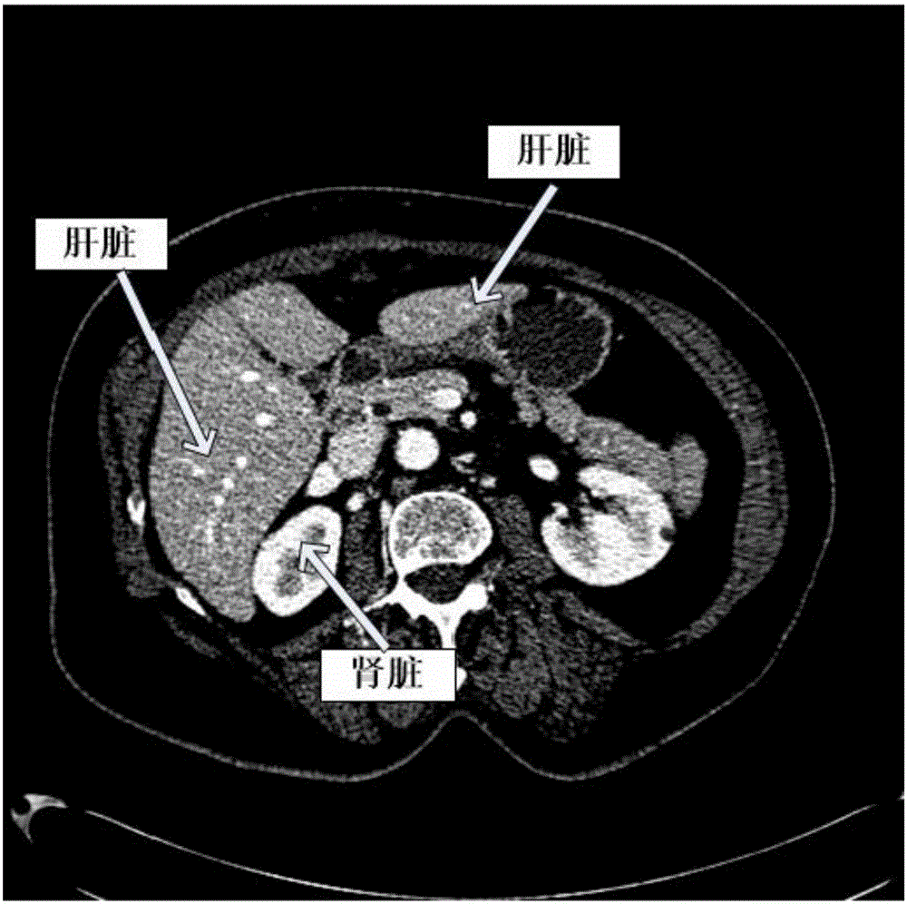
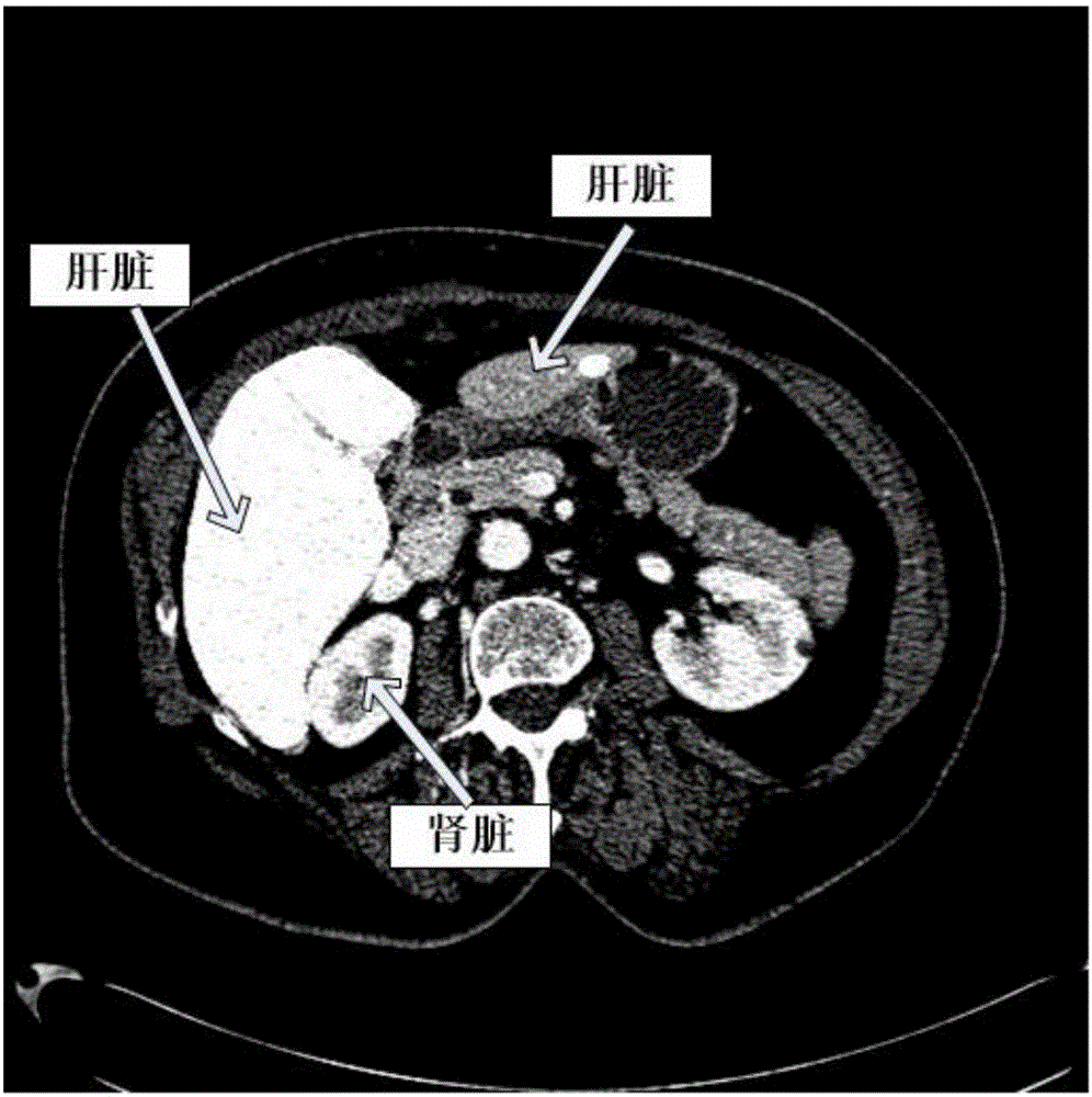
[0051] Such as figure 1 As shown, the automatic three-dimensional liver segmentation method based on local prior information and convex optimization is used to segment the liver in the computed tomography angiography image. The specific steps are:

[0052] 1. The size of the input liver CTA scan image I is 512×512×245, adjust the window width and window level to make the liver gray range between 0 and 255. The image is tested in the trained convolutional neural network, and a probability map L with the same size as the original image is obtained. In the probability map, the probability L(x) of each point belonging to the liver in a given image is in the range of [0.1, 0...


PUM


Abstract

The invention relates to medical image processing, and aims to provide a fully-automatic three-dimensional liver segmentation method based on local apriori information and convex optimization. The fully-automatic three-dimensional liver segmentation method based on the local apriori information and the convex optimization comprises the following steps: processing abdominal liver CTA volume data by utilizing a trained three-dimensional convolutional neural network, and then obtaining an aprior probability graph of a liver; obtaining an initial region of the liver from the aprior probability graph of the liver; determining probabilities of various pixel points, belonging to a foreground liver and a background, in an image; optimizing a new energy model by utilizing a convex optimization technology, and segmenting the liver; and performing post-processing, and then obtaining a contour of the liver. The method obtains a segment result. The method can overcome problems of under segmentation and inaccurate boundaries, which exist in an original liver segmentation by utilizing a convolutional neural network, well, and then can obtain an accurate segmentation result.

Description

technical field [0001] The invention relates to the field of medical image processing, in particular to a fully automatic three-dimensional liver segmentation method based on local prior information and convex optimization. Background technique [0002] At present, liver disease is a disease with a relatively high incidence rate clinically, which directly threatens people's lives, so the accurate diagnosis of liver disease has important medical significance. Clinically, doctors often use a CT machine, that is, a computerized tomography machine, to obtain a series of planar grayscale tomographic images of the liver, and to judge the lesion location, characteristics, size, and surrounding tissue of the lesion by continuously viewing these images. relationship between etc. The extraction and quantitative analysis of the liver play a key role in accurately diagnosing liver diseases and formulating appropriate surgical plans. Clinically, the extraction of the liver is often out...

Claims


Application Information

Patent Timeline
IPC IPC(8): G06T7/00
CPCG06T7/0012G06T2207/10081G06T2207/20084G06T2207/30056
Inventor 孔德兴胡佩君吴法
Owner ZHEJIANG DE IMAGE SOLUTIONS CO LTD
Why Eureka
Try Eureka