Blood cell and biochemical component analysis instrument and method

A component analysis and blood cell technology, which is applied in the field of medical inspection and testing, can solve the problems that the detection mode is not suitable for rapid detection, the sample coding information is prone to errors, and the detection items are single, so as to save detection time and eliminate biochemical detection results. difference, the effect of accurate test results

Active Publication Date: 2016-11-16
SINNOWA MEDICAL SCI & TECH
View PDF7 Cites 8 Cited by
  • Summary
  • Abstract
  • Description
  • Claims
  • Application Information

AI Technical Summary

Problems solved by technology

[0003] Existing blood cell testing and biochemical testing are generally performed on blood analyzers and biochemical analyzers respectively. Therefore, two blood samples and two different instruments must be prepared for testing. Error, this detection mode is especially not suitable for the needs of rapid detection in outpatient clinics and emergency departments
Moreover, the existing instruments that can perform blood routine and biochemical testing have a single test item, a complex structure, and a cumbersome process

Method used


Image

  • Blood cell and biochemical component analysis instrument and method
  • Blood cell and biochemical component analysis instrument and method
  • Blood cell and biochemical component analysis instrument and method

Examples


Embodiment 1

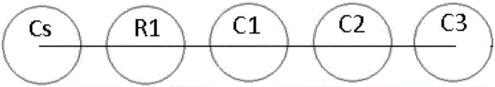
[0097] In this embodiment, four detection cups are taken as an example to detect blood routine and single-reagent biochemical items.

[0098] like figure 2 As shown, the first detection cup C1 is provided with a first particle count detection device D4, a first transmitted light detection device and a first biochemical detection device, the first biochemical detection device includes a first scattered light detection device; the first transmitted light detection device It includes a first transmitted light light source L1; the first scattered light detection device includes a first scattered light light source L4, and the first scattered light detection device shares a first detector D1 with the first transmitted light detection device.

[0099] The second detection cup C2 is provided with a second particle counting detection device D5 and a second biochemical detection device, and the second biochemical detection device includes a second transmitted light detection device an...

Embodiment 2

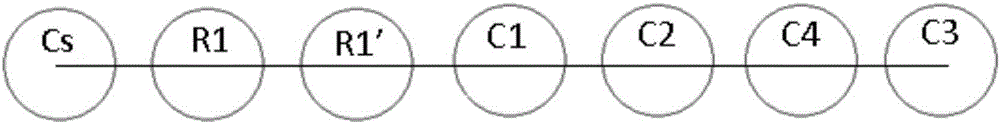
[0111] combine figure 2 and Figure 4 The difference between the analyzer of this embodiment and that of Embodiment 1 is that in this embodiment, blood routine and dual-reagent biochemical items are detected.

[0112] The sampling needle N1 moves to the first biochemical reagent position R1, and the sampling needle N1 penetrates into the first biochemical reagent, and with the cooperation of the first liquid extraction device S1, the first valve V1 and the second valve V2, draws the quantitative first biochemical reagent Afterwards, the sampling needle N1 moves to the first detection cup C1, the first biochemical reagent is added to the first detection cup C1, the eleventh valve V11 and the ninth valve V9 in the mixing module are opened, the tenth valve V10, the The twenty-second valve V22, the twenty-third valve V23 and the twelfth valve V12 are closed, and the air pump P1 blows air, driving the gas along the pipeline into the bottom of the first detection cup C1, and mixin...

Embodiment 3

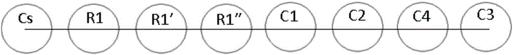
[0114] The difference between this embodiment and Embodiment 1 and Embodiment 2 is that the first transmitted light source L1 is turned on, the first scattered light source L4 is turned off, and the first detector D1 detects the transmitted light signal to obtain the detection results of biochemical items.


PUM


Abstract

The invention discloses a blood cell and biochemical component analysis instrument and method. The analysis instrument comprises a transfer module, a first hemolytic agent adding module, a second hemolytic agent adding module, a third hemolytic agent adding module, a diluent adding module, a cleaning agent adding module, a cleaning module, an even mixing module, a control module, a first detection cup, a second detection cup, a third detection cup and a fourth detection cup. The blood cell and biochemical component analysis instrument can achieve rapid completion of conventional and biochemical item detection of blood cells in one, makes detection functions of the instrument stronger, makes the sample usage amount smaller, makes the detection efficiency higher and provides more comprehensive information for clinic disease diagnosis.

Description

technical field [0001] The invention belongs to the technical field of medical inspection and detection, in particular to a blood cell and biochemical component analyzer and method. Background technique [0002] In medical clinical diagnosis, it is often necessary for the laboratory to obtain the patient's blood blood cell test information and biochemical test information at the same time. The comprehensive test information provided by the laboratory can provide more complete guidance information for clinical disease diagnosis and treatment. [0003] Existing blood cell testing and biochemical testing are generally carried out on blood analyzers and biochemical analyzers respectively. Therefore, two blood samples and two different instruments must be prepared for testing. Wrong, this detection mode is especially not suitable for the needs of rapid detection in outpatient clinics and emergency departments. Moreover, the existing instruments that can perform blood routine and...

Claims


Application Information

Patent Timeline
Patent Type & Authority Applications(China)
IPC IPC(8): G01N33/50G01N35/00
CPCG01N33/48G01N35/02G01N33/50G01N35/00584
Inventor 徐新董自权曹宁宋成桥周强郭敏
Owner SINNOWA MEDICAL SCI & TECH
Why Eureka
Try Eureka