Full-automatic fundus camera

A fully automatic, camera-based technology, applied in the field of medical imaging, can solve the problems that ordinary people cannot use it normally, and achieve the effect of universal screening and remote diagnosis.

Inactive Publication Date: 2017-03-15
SUZHOU MICROCLEAR MEDICAL INSTR
View PDF5 Cites 11 Cited by
  • Summary
  • Abstract
  • Description
  • Claims
  • Application Information

AI Technical Summary

Problems solved by technology

[0003] Since the fundus camera is a professional medical device, it needs professional personnel to operate and interpret it, and ordinary people cannot use it normally

Method used


Image

  • Full-automatic fundus camera

Examples


Embodiment Construction

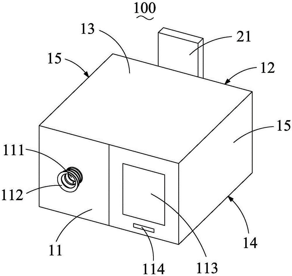
[0019] see figure 1 As shown, the fully automatic fundus camera 100 of the present invention includes a housing 10, an imaging module housed in the housing 10, an illumination module, a fixation module and a three-degree-of-freedom movement module, a pupil positioning unit, an autofocus unit, and a communication module. , a height adjustment module, a printing module and a scanning module.

[0020] see figure 1 As shown, the housing 10 is roughly in the shape of a cuboid, including a front wall 11, a rear wall 12 opposite to the front wall 11, a top wall 13 perpendicular to the front wall 11, and a top wall 13 perpendicular to the top wall 13. The oppositely arranged bottom wall 14 and a pair of side walls 15 perpendicular to the front wall 11 and the top wall 13, the front wall 11, the rear wall 12, the top wall 13, the bottom wall 14 and the side walls 15 jointly surround A storage space is formed to accommodate the imaging module, illumination module, vision fixation modu...


PUM


Abstract

The invention discloses a full-automatic fundus camera. The full-automatic fundus camera comprises a shell, an image-forming module, a three degrees-of-freedom motion module, a pupil location unit and an automatic focusing unit. Compared with the prior art, a professional fundus retinal image can be automatically shot as long as a photographed person focuses on an observation window, so that a professional doctor can conduct a remote diagnosis and then push the diagnosis information to the photographed person, and thus the fundus camera is made likely to enter a grassroot health-center and a medical institution in a remote area, and whole-people popularization and screening can be achieved.

Description

technical field [0001] The invention relates to the field of medical imaging, in particular to a fully automatic fundus camera. Background technique [0002] The fundus camera belongs to the field of medical imaging and is used to obtain retinal images of the human eye so that medical personnel can check fundus diseases or assist medical personnel to judge the condition of other organs. Because the blood vessels of the fundus are the only blood vessels that can be directly observed through the body surface, medical personnel can check whether there are lesions in the optic nerve, retina, choroid and refractive medium of the fundus through the fundus camera, and can also use the fundus camera to assist other systems. Diagnosis and judgment of diseases, such as detecting cerebral infarction, cerebral hemorrhage, cerebral arteriosclerosis, brain tumors, diabetes, kidney disease, hypertension, retinopathy of prematurity, glaucoma, age-related macular degeneration, etc. by screen...

Claims


Application Information

Patent Timeline
Patent Type & Authority Applications(China)
IPC IPC(8): A61B3/15A61B3/12
CPCA61B3/152A61B3/0008A61B3/12A61B3/154
Inventor 李超宏
Owner SUZHOU MICROCLEAR MEDICAL INSTR
Why Eureka
Try Eureka