SVM (support vector machine)-based medical image blood vessel recognition method

A recognition method and medical image technology, applied in the computer field, can solve the problem of not being able to extract blood vessels too accurately, and achieve the effects of improving accuracy, preserving blood vessel bifurcation, and enhancing blood vessel network.

Inactive Publication Date: 2017-03-22
BEIJING UNIV OF TECH
View PDF6 Cites 32 Cited by
  • Summary
  • Abstract
  • Description
  • Claims
  • Application Information

AI Technical Summary

Problems solved by technology

[0007] However, only through the existing methods described above, the blood vessels cannot be extracted very accurately, especially the fundus blood vessels with blurred images such as cataracts, which make

Method used


Image

  • SVM (support vector machine)-based medical image blood vessel recognition method

Examples


Embodiment Construction

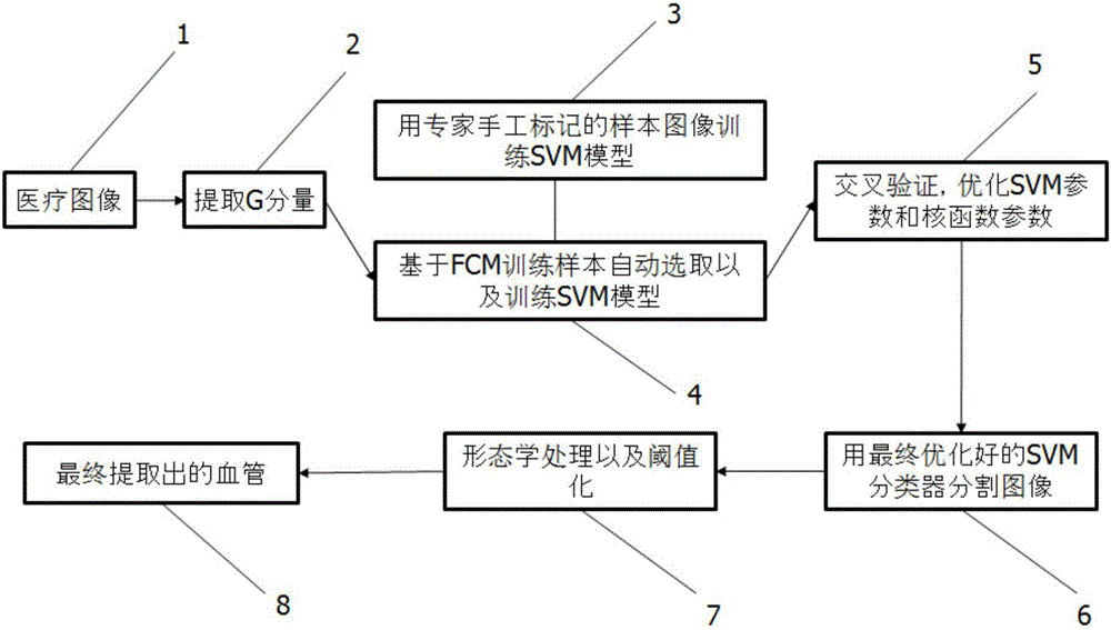
[0029] Features and illustrative examples of various aspects of the invention are described in detail below. The software used to realize this blood vessel extraction method can be Matlap or OpenCV. Both development tools have great image manipulation capabilities.

[0030]An embodiment of the present invention is an SVM-based blood vessel recognition method in a medical image, which specifically includes two parts: first, SVM is used to initially segment blood vessels, and then morphological operations and thresholding are used for processing. Among them, the SVM segmentation of blood vessels is actually to divide the blood vessels into foreground and background (ie, blood vessels and non-vascular) parts by the SVM classifier trained by the training set. Among them, the training set samples include samples automatically selected by FCM and samples manually divided by experts. Morphological operation and thresholding processing include three steps: grayscale inversion, high-...


PUM


Abstract

The invention discloses an SVM (support vector machine)-based medical image blood vessel recognition method. The method includes the following steps that: since an SVM classifier is obtained after cross validation and optimization are performed on an SVM model trained by samples automatically selected by an FCM by using an SVM model which is trained by samples obtained through the manual segmentation of experts, segmentation results are more accurate; the SVM is adopted to segment a blood vessel, namely, pixels are divided into a foreground (blood vessels) and a background (other parts), and the blood vessel part is extracted out; morphological processing and thresholding are performed, and therefore, a blood vessel network can be enhanced, the branches and intersections of the blood vessels are reserved; and an obtained image is converted into a binary image, so that blood vessel distribution can be reflected more directly. According to the method of the invention, the FCM, the SVM and the morphological image processing are combined together, so that a recognition effect is better.

Description

technical field [0001] The invention belongs to the field of computers, and proposes an SVM-based blood vessel recognition method in medical images, which can well extract blood vessel networks in medical images, especially blood vessels in fundus images. Background technique [0002] In medical diagnosis and medical research, blood vessels are very important biological tissues. When many organs are diseased, the blood vessels will appear abnormal. Such as retinal blood vessels in the eyes, coronary arteries in the heart, small blood vessels in the lungs, etc. Among them, the retina is the only relatively deep blood vessel in the human body that can be directly observed in a non-invasive way, and the retinal blood vessel is the most important anatomical structure visible in fundus images, and changes in its structural characteristics directly reflect various lesions in the body. Retinal fundus images can also be used as an important basis for judging cataract and other dise...

Claims


Application Information

Patent Timeline
IPC IPC(8): G06T7/00G06T7/136
CPCG06T7/0012G06T2207/20081G06T2207/30041G06T2207/30101
Inventor 胡启东李建强张苓琳韩赫
Owner BEIJING UNIV OF TECH
Why Eureka
Try Eureka