Medical image automatic segmentation method based on deep learning

A technology for automatic segmentation of medical images, applied in the field of medical image processing, can solve problems such as difficult detection of small targets and blurred boundaries of segmented targets

Pending Publication Date: 2022-02-18
HUNAN UNIV OF SCI & TECH
View PDF0 Cites 7 Cited by
  • Summary
  • Abstract
  • Description
  • Claims
  • Application Information

AI Technical Summary

Problems solved by technology

[0005] Aiming at the shortcomings and deficiencies of the prior art, the present invention integrates the residual module and the attention mechanism into the construction of a U-shaped deep convolutional neural network, aiming to provide a method for automatically segmenting medical image target regions based on deep learning, and to solve the problem of medical image The boundary of middle segmentation target is blurred, and it is difficult to detect small targets, so as to improve the accuracy and efficiency of computer-aided diagnosis

Method used


Image

  • Medical image automatic segmentation method based on deep learning
  • Medical image automatic segmentation method based on deep learning
  • Medical image automatic segmentation method based on deep learning

Examples


Embodiment 1

[0030] A method for automatic segmentation of medical images based on deep learning, the specific implementation steps are as follows:

[0031] (1) Obtain the patient's original medical image and the artificial segmentation results of the target area in the image from the medical image public data set, and construct a training data set;

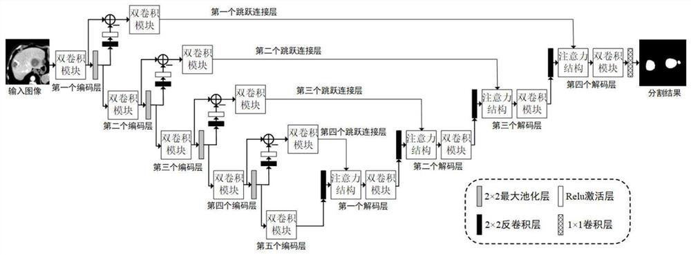
[0032] (2) Build as figure 1 The U-shaped deep convolutional network that fuses the residual module and the attention mechanism shown is called RA-Unet, including:

[0033](2-a) U-shaped network is used as the backbone network. The backbone network consists of five encoding layers, four skip connection layers, four decoding layers and one 1×1 convolutional layer. The output of the first encoding layer Not only as the input of the second encoding layer, but also connected to the fourth decoding layer through the first skip connection layer as the input of the decoding layer; the output of the second encoding layer is not only used as the inpu...

Embodiment 2

[0048] The method in Example 1 was used to conduct experiments on the MICCAI 2017 pancreas public segmentation dataset. There are two segmentation categories: background, pancreas organ. The computer environment of this experiment: the operating system is Windows 10; an NVIDIA RTX308010G GPU; the software platform is: Python3 and PyTorch 1.6.

[0049] In this embodiment, the original CT images of 80 cases and the corresponding artificial segmentation results of the pancreas are selected from the pancreas public segmentation data set as the training data set, and the original CT images of 20 cases are selected as the test data set. In the experiment, the average Dice coefficient was used as the evaluation standard, and the segmentation method of the present invention achieved a Dice coefficient of 85.12% on the task of pancreas segmentation, and obtained a relatively high segmentation accuracy. The partial image segmentation results of the test set are as follows: Figure 4 (...

Embodiment 3

[0051] Using the method in Example 1, the kidney organ segmentation experiment was performed on the MICCAI 2017 kidney public segmentation dataset. There are two segmentation categories: background, kidney organ. The computer environment of this experiment: the operating system is Windows 10; an NVIDIA RTX3080 10G GPU; the software platform is: Python3 and PyTorch 1.6.

[0052] In this embodiment, the original CT images of 126 cases and the corresponding artificial kidney segmentation results are selected from the kidney public segmentation data set as the training data set, and the CT images of 84 cases are selected as the test data set. In the experiment, the average Dice coefficient was used as the evaluation standard, and the segmentation method of the present invention achieved a Dice coefficient of 91.12% on the pancreas segmentation task. The partial image segmentation results of the test set are as follows: Figure 5 (a)~ Figure 5 As shown in (d), the area shown by...


PUM


Abstract

The invention discloses a medical image automatic segmentation method based on deep learning, and the method specifically comprises the steps: (1) obtaining a manual segmentation result of an original medical image of a patient and a target region, and constructing a training data set related to a segmentation task; (2) designing a deep U-shaped convolutional neural network model fusing a residual module and an attention mechanism; (3) constructing a mixed loss function by using Dice and binary cross entropy; (4) performing network training by using the training data set; and (5) segmenting a target area in the test image by using the trained network. The invention relates to an end-to-end medical image full-automatic segmentation method, which can effectively solve the problems of fuzzy segmentation boundary, low small target detection rate, unstable training process, difficulty in convergence and the like in the current deep learning image segmentation field.

Description

technical field [0001] The invention belongs to the technical field of medical image processing, and in particular relates to an automatic medical image segmentation method based on deep learning. Background technique [0002] With the rapid development of medical imaging technology, more doctors will use medical imaging technology to diagnose patients' conditions and propose treatment plans. Currently common medical imaging techniques include: Computed Tomography (CT), Magnetic Resonance Imaging (MRI) and so on. Medical image processing is an important technical means to assist doctors in understanding patients' conditions. Imaging technology can help doctors understand the lesions in patients more intuitively and make accurate and effective diagnoses. [0003] Medical image segmentation refers to the separation of diseased areas, endangered organs, and radiotherapy target areas in the original medical image. It is an important part of medical image processing. play a vit...

Claims


Application Information

Patent Timeline
Patent Type & Authority Applications(China)
IPC IPC(8): G06T7/00G06T7/136G06K9/62G06N3/04G06N3/08G06V10/82G06V10/774
CPCG06T7/0012G06T7/136G06N3/08G06T2207/10081G06T2207/30096G06N3/048G06N3/045G06F18/214
Inventor 廖苗邸拴虎杨文瀚赵于前杨振曾业战
Owner HUNAN UNIV OF SCI & TECH
Why Eureka
Try Eureka