
Correction method and system for ultrasonic three-dimensional image

A three-dimensional image, three-dimensional technology, applied in image enhancement, image analysis, image data processing and other directions, can solve the problems of different images, unfavorable observation and diagnosis, etc., to achieve the effect of improving convenience and convenient comparison and viewing

Active Publication Date: 2019-11-01
SONOSCAPE MEDICAL CORP
View PDF4 Cites 0 Cited by
  • Summary
  • Abstract
  • Description
  • Claims
  • Application Information

AI Technical Summary

Problems solved by technology

[0003] Due to the position of the probe of the ultrasound imaging system, the movement of the fetus or tissues and organs, the orientation of the target area of ​​interest may change easily, and the orientation of the images collected in different periods may be different, which is not conducive to observation and diagnosis

Method used


Image

  • Correction method and system for ultrasonic three-dimensional image
  • Correction method and system for ultrasonic three-dimensional image
  • Correction method and system for ultrasonic three-dimensional image

Examples


Embodiment

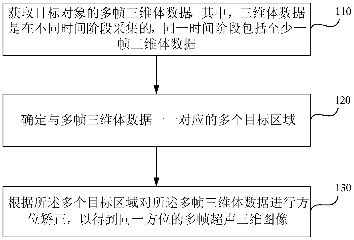
[0061] figure 1 A flow chart of a method for correcting an ultrasonic three-dimensional image provided by an embodiment of the present invention, the method is applicable to correcting multiple frames of three-dimensional volume data based on time sequence with a fixed orientation, and can be performed by a correction system for an ultrasonic three-dimensional image . The system can be realized by means of hardware and / or software. The method specifically includes the following steps:

[0062] Step 110, acquiring multiple frames of 3D volume data of the target object, wherein the 3D volume data is collected at different time periods, and the same time period includes at least one frame of 3D volume data.

[0063] The target object is the same tissue and organ of the same object to be examined or the fetus in the abdomen of the same pregnant object. The multiple frames of three-dimensional volume data are collected in different time periods, and the time periods include at l...


PUM


Abstract

The embodiment of the invention discloses a method and a system for correcting an ultrasonic three-dimensional image. The method comprises steps: multiple frames of three-dimensional body data of a target object are acquired, wherein the three-dimensional body data are acquired in different time periods, and at least one frame of three-dimensional body data are included in the same time period; multiple target areas in one-to-one correspondence with the multiple frames of three-dimensional body data are determined; and direction correction is carried out on the multiple frames of three-dimensional body data according to the multiple target areas to obtain multiple frames of ultrasonic three-dimensional images in the same direction. According to the method of correcting the ultrasonic three-dimensional image disclosed by the embodiment of the invention, the multiple frames of three-dimensional body data in different time periods and in different directions of the target object can be corrected to be in the same direction, comparison and lookup by the user are facilitated, and the convenience is thus improved.

Description

technical field [0001] Embodiments of the present invention relate to the technical field of ultrasonic imaging, and in particular to a method and system for correcting an ultrasonic three-dimensional image. Background technique [0002] With the development of medical technology, ultrasonic four-dimensional imaging has been widely used in the medical industry. Ultrasound four-dimensional imaging is mainly used to view the anatomical structure of tissues, or for obstetric ultrasound examination, such as imaging embryos or fetuses in the mother's uterus, displaying real-time dynamic moving images of fetuses, or displaying real-time moving images of human internal organs. Three-dimensional images are static pictures, and four-dimensional images are based on dynamic images within a certain period of time. [0003] Due to the position of the probe of the ultrasound imaging system, the movement of the fetus or tissues and organs, the orientation of the target area of ​​interest ...

Claims


Application Information

Patent Timeline
Patent Type & Authority Patents(China)
IPC IPC(8): G06T5/50
CPCG06T5/50G06T2207/10021G06T2207/10136G06T2207/30044
Inventor 艾金钦刘旭江孙慧黄柳倩喻美媛唐艳红
Owner SONOSCAPE MEDICAL CORP
Learn More