Method for extracting brain glioma region from brain nuclear magnetic resonance image

A technology for nuclear magnetic resonance images and brain gliomas, applied in the field of medical images, to achieve fast convergence, wide application value and prospects, and high stability

Active Publication Date: 2017-05-31
NANJING AUDIT UNIV
View PDF5 Cites 20 Cited by
  • Summary
  • Abstract
  • Description
  • Claims
  • Application Information

AI Technical Summary

Problems solved by technology

[0005] The technical problem to be solved by the present invention is to provide a method for extracting brain glioma regions in brain MRI images that can effectively improve the accuracy of brain glioma region extraction for degraded problems such as noise and bias field

Method used


Image

  • Method for extracting brain glioma region from brain nuclear magnetic resonance image
  • Method for extracting brain glioma region from brain nuclear magnetic resonance image
  • Method for extracting brain glioma region from brain nuclear magnetic resonance image

Examples


Embodiment Construction

[0069] The specific implementation manners of the present invention will be further described in detail below in conjunction with the accompanying drawings.

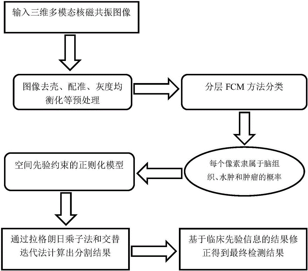
[0070] Such as figure 1 As shown, the present invention designs a method for extracting glioma regions in brain MRI images. In practical applications, it specifically includes the following steps:

[0071] Step A. Preprocessing and fusion operations are performed on each target multimodal brain MRI image to obtain a multimodal fusion MRI image of the target brain.

[0072] Wherein, step A includes the following steps:

[0073] Step A1. Use the N4ITK method to perform debiasing field operations on each target multi-modal brain MRI image, and update each target multi-modal brain MRI image.

[0074] Step A2. Use the Non-rigid-based image registration method to register each target multimodal brain MRI image according to the reference brain standard weight image, and update each target multimodal brain MRI image image.

...


PUM


Abstract

The invention relates to a method for extracting a brain glioma region from a brain nuclear magnetic resonance image. The method can realize accurate segmentation of the brain glioma region. Compared with previous solution, the method provided by the invention has the advantages that a brain glioma region extraction general model is built by adopting a combined layering FCM method and a total variation regularization method, the characteristics of an MRI sequence image corresponding to each pixel are fully considered, the spatial information of each pixel is also introduced, the characteristics of the MRI sequence image and the spatial information are organically combined and co-act on the segmentation of the brain glioma region, and the brain glioma extraction accuracy is improved.

Description

technical field [0001] The invention relates to a method for extracting a brain glioma region in a brain nuclear magnetic resonance image, and belongs to the technical field of medical images. Background technique [0002] Tumors are the products of the wanton growth of cancer cells in the body. They have different shapes and presentation methods, and the specific treatment methods are also different. At present, brain tumors have become a common disease all over the world. In 2015, there were nearly 2,300 new brain cancer patients in the United States. There are many types of brain tumors, among which glioma is a common and highly malignant brain tumor with high mortality and disability rates. According to clinical knowledge, gliomas can be divided into high-grade gliomas and low-grade gliomas according to their malignancy. In order to accurately diagnose glioma, multimodal MRI images are often used in clinical detection of glioma. Currently commonly used modalities incl...

Claims


Application Information

Patent Timeline
Patent Type & Authority Applications(China)
IPC IPC(8): G06T7/11G06T7/187G06T7/00
CPCG06T7/0012G06T2207/10088G06T2207/30016G06T2207/30096
Inventor 杨章静詹天明万鸣华张辉杨国为
Owner NANJING AUDIT UNIV
Why Eureka
Try Eureka