Application of peripheral free exosomes to preparation of liquid-state biopsy tumor diagnosis reagents

A tumor diagnosis and exosome technology, applied in measurement devices, instruments, scientific instruments, etc., can solve the problems that ctDNA detection technology is not suitable for clinical application, the detection effect of early tumor patients is not very good, and the variation of ctDNA quantity is large. , to achieve far-reaching clinical significance and promotion, improve diagnostic specificity, easier access and enrichment effects

Active Publication Date: 2017-06-09
杭州联川基因诊断技术有限公司
View PDF6 Cites 9 Cited by
  • Summary
  • Abstract
  • Description
  • Claims
  • Application Information

AI Technical Summary

Benefits of technology

This patented technology allows researchers to identify different types of particles (microparticles), called extracellular microbons(EMB). These particles have been found to circulate around within our body throughout its lifetimes without causing harmful side effects. They may contain important materials like DNA fragments, proteins, carbohydrates, lipids, amino acids, peptides, nucleate polysaccharidases, etc., allowing them to actively transport these small objects towards target organs through passages inside the body. By measuring this gene activity levels at multiple locations along the entire organism's surface, we aim to provide an effective way to monitor diseased individuals who develop signs caused by abnormalities associated with inflammatory responses.

Problems solved by technology

Technologies aim at developing an improved way to accurately identify and measure minute amounts of cell material called Circulated Tissue Gastrointestinal Veins (CNT). These tiny tubules contain various types of gas bags containing air instead of water, making them ideal candidates for non-destructive testing without causing any harm caused by other substances like drugs or radiation exposures. Current technologies require multiple steps involving sampling and analysis before accurate results may become available. Additionally, they lack precision when extracting rare nucleic acid sequences specifically targeting certain areas within the sample.

Method used


Image

  • Application of peripheral free exosomes to preparation of liquid-state biopsy tumor diagnosis reagents
  • Application of peripheral free exosomes to preparation of liquid-state biopsy tumor diagnosis reagents
  • Application of peripheral free exosomes to preparation of liquid-state biopsy tumor diagnosis reagents

Examples


Embodiment 1

[0046] Embodiment 1 prepares detection kit

[0047] Take 1ml of plasma as the object:

[0048] (1) 200 μl total exosome isolation reagent (Total Exosome Isolation, Invitrogen);

[0049] (2) 20 μl of EpCAM separation magnetic beads (EpCAM beads, Invitrogen), 3 ml of 1×PBS;

[0050] (3) Elisa detection kit, including:

[0051] 100 μl 1:5000 diluted CD9 primary antibody (proteintech) (original concentration 56 μg / 150 μl, concentration after 1:5000 dilution is 0.0000746 μg / μl);

[0052] 100 μl 1:10000 diluted HRP enzyme-labeled secondary antibody (sigma) (original concentration 0.4 μg / μl, concentration after 1:10000 dilution is 0.00004 μg / μl);

[0053] High-binding microtiter plate; 200μl PBST containing 5% skimmed milk as blocking solution; 100μl TMB chromogenic solution; 100μl stop solution; 200μl 1×PBS; 12*200μl 1×PBST.

[0054] Wherein, PBS refers to phosphate buffered saline solution, PBST contains Tween-20 in PBS, and the mass percentage of Tween-20 in the whole PBST sol...

Embodiment 2

[0055] Example 2 Using the kit of Example 1 to detect the expression of CD9 in peripheral free exosomes of patients with renal cancer

[0056] (1) Extraction and purification of tumor exosomes

[0057] ①With the patient’s informed and ethical consent, collect 10ml of preoperative blood samples from tumor patients (pathological biopsy has confirmed renal clear cell carcinoma) in blood collection tubes, centrifuge at 4°C and 300g for 10 minutes, separate To remove the cells in the blood, and then obtain 1ml of the upper layer of plasma.

[0058] ② Centrifuge the plasma at 4°C and 10,000g for 30 minutes, separate to remove organelles and other impurities, and obtain a supernatant.

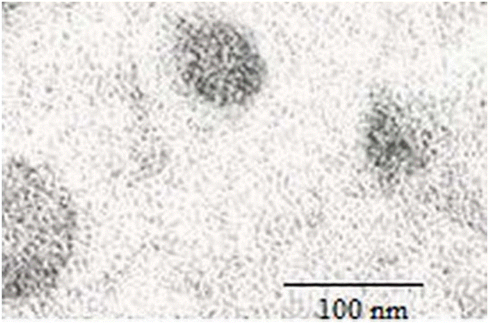
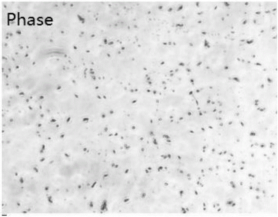
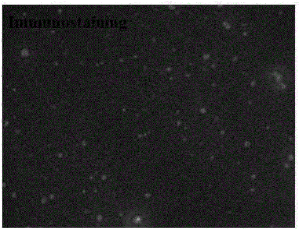
[0059] ③Put the supernatant into a new centrifuge tube, and add 200 μl of exosome extract (TotalExosome Isolation, Invitrogen) into it to mix, shake evenly, and incubate at 4°C overnight (6-16 hours are acceptable), then, incubate The final mixture was centrifuged at 4° C. and 10,000 g for 1 hour, a...

Embodiment 3

[0097] Example 3 Using the kit of Example 1 to detect the expression of CD9 in peripheral free exosomes of patients with lung cancer

[0098] Using basically the same method as step (1) in Example 2, the collected plasma (1ml) of 30 cases of lung cancer patients and 30 cases of healthy human plasma (1ml) were used to extract and purify tumor exosomes to obtain magnetic beads Bound Epcam-positive (tumor cell-derived) exosome. That is: the plasma sample in (1) is a preoperative plasma sample of a lung cancer patient, and other steps are exactly the same as in Example 2. The method for obtaining the plasma sample is also the same as step (1) ① in Example 2.

[0099] Using the method basically the same as step (2) in Example 2, the ELisa detection of peripheral free tumor exosomes was carried out. The expression of CD9 was detected on the Epcam-positive (tumor cell-derived) exosomes obtained above in Example 3, and analyzed and compared by scatter plots. A scatterplot such as ...


PUM


Abstract

The invention provides application of peripheral free exosomes to preparation of liquid-state biopsy tumor diagnosis reagents. Specific markers CD9 of the free exosomes in peripheral blood of tumor patients are remarkably higher than specific markers CD9 of exosomes in healthy individuals of a control group in expression level, thereby being high in specificity and sensitivity when serving as tumor diagnosis markers. The invention further provides a kit for tumor diagnosis. The kit for tumor diagnosis comprises the reagents for detecting the specific markers of the free exosomes in the peripheral blood, wherein the reagents include (1) a total exosome isolation reagent for isolating total exosomes from the peripheral blood, (2), an EpCAM isolating magnetic bead for purifying tumor-derived exosomes from the total exosomes, (3) an Elisa detection reagent for detecting expression of specific markers in the tumor-derived exosomes. The kit can be applied to tumor diagnosis and monitoring, in particular to screening of tumor high-risk groups, early noninvasive diagnosis or prognosis evaluation of the patients.

Description


Claims


Application Information

Patent Timeline
Owner 杭州联川基因诊断技术有限公司
Why Eureka
Try Eureka