Method for detecting carcinogenesis in the uterine cervix

A cervical cancer and protein technology, applied in measuring devices, biological tests, material inspection products, etc., to increase diagnostic accuracy, reduce over-diagnosis and over-processing, and increase overall sensitivity and specificity

Inactive Publication Date: 2017-08-01
文森佐麦卡里尼 +1
View PDF11 Cites 0 Cited by
  • Summary
  • Abstract
  • Description
  • Claims
  • Application Information

AI Technical Summary

Problems solved by technology

[0016] To date, however, there are no biomarkers indicating irreversible transformation of premalignant lesions to cervical cancer (KooYJ. et al., Dual immunostaining of cervical cytology specimens with atypical squamous cells for p16 / Ki-67 does not exclude the existence of a high-grade squamous intraepithelial lesion.Virchows Arch.2013Oct 1; Pac-chiarotti A. et al., Prognostic value of p16-INK4A protein in women with negative or CIN1 histology result: A follow-up study.Int J Cancer.2014,134,897-904)

Method used


Image

  • Method for detecting carcinogenesis in the uterine cervix
  • Method for detecting carcinogenesis in the uterine cervix
  • Method for detecting carcinogenesis in the uterine cervix

Examples


Embodiment Construction

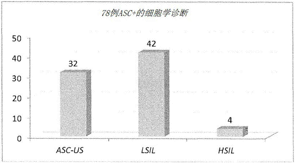
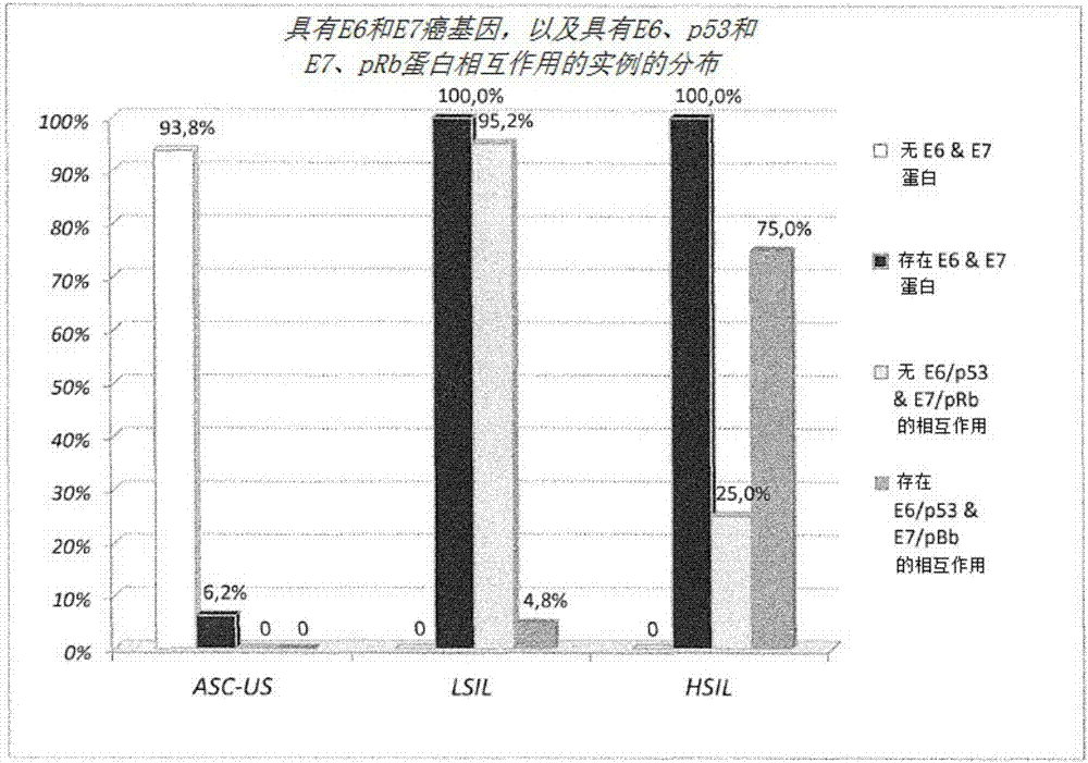
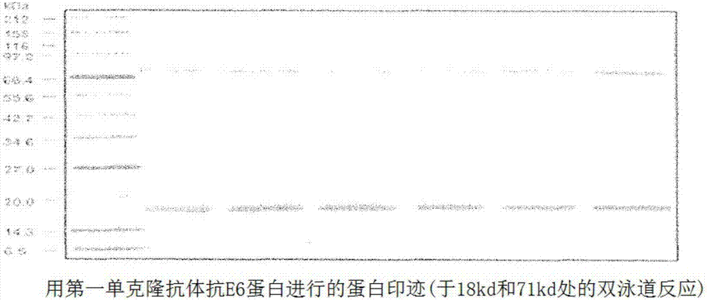
[0028] According to its broadest embodiment, therefore, the present invention provides in particular a method for detecting neoplastic transformation caused by human papillomavirus (HPV) in women undergoing cervical cancer screening, said method comprising Cell samples from the squamous-columnar junction of the cervical epithelium of the examined patients were detected for the presence of the protein complex E6 / p53 consisting of protein E6 and protein p53 and / or the protein complex E7 / p53 consisting of protein E7 and protein pRb The presence of pRb, in which at least one of the two protein complexes is detected, indicates that, in the patient examined, the carcinogenesis has become irreversible, preferably by means of an antibody against the protein in the analytical technique of Western blot or sandwich ELISA The detection is carried out using a combination of E6 and an antibody against the protein E7 and preferably an antibody against the protein p53 and an antibody against t...


PUM

PropertyMeasurementUnit
molecular weightaaaaaaaaaa

Abstract

The invention concerns some methods for diagnosing cancer of the uterine cervix (or extra-uterine) which integrate or surrogate the field of action of cytology with that of molecular biology, and provide a system for diagnosis, prognosis and management of HPV-induced cervical lesions (or extra-uterine) which exploit, in particular, the analytical methods of Western Blot and Sandwich ELISA in order to detect those cases where the transformation towards a neoplastic lesion has become irreversible. The methods consist in detecting, in samples of cells taken from the squamous-columnar junction of the uterine cervix of a patient under examination, the proteins encoded by viral oncogenes E6 and E7 and those encoded by the tumor suppressor genes of the host cell p53 and pRB, and in detecting by Western Blot and / or by Sandwich ELISA the possible interaction between proteins E6 and p53, and between proteins E7 and pRb, as an index of irreversible transformation towards a neoplastic lesion.

Description

[0001] describe [0002] field of invention [0003] The present invention relates to methods of detecting cancerous changes in the cervix. More specifically, the present invention relates to procedures for diagnosing cervical cancer that integrate or replace the fields of action of cytology and molecular biology, and provide systems for the diagnosis, prognosis, and management of HPV-induced cervical lesions, It employs very sensitive and specific analytical methods, such as Western blot and sandwich ELISA, in order to detect those cases where the transformation towards neoplastic lesions has become irreversible. [0004] Background of the invention [0005] The link between human papillomavirus (HPV) infection and cancer has sparked continued interest among researchers. HPV infects the basal cells of the squamous epithelium and induces the formation of benign lesions and / or in some cases aggressive malignant tumors. HPV virions consist of a protein coat (capsid) surroundin...

Claims


Application Information

Patent Timeline
Patent Type & Authority Applications(China)
IPC IPC(8): G01N33/574
CPCG01N33/57411G01N2800/56G01N2333/025G16H50/20G16B30/00G01N33/574G01N27/44747G01N33/5091G01N33/52G01N33/5748G01N33/581
Inventor 文森佐·麦卡里尼朱塞佩·迪贝内德托
Owner 文森佐麦卡里尼
Why Eureka
Try Eureka