
Reconstruction method and device of medical scanning image and medical imaging system

A scanning image and image reconstruction technology, applied in the field of medical scanning image processing, can solve the problems of resolution degradation in the Z-axis direction, reconstruction layer thickness broadening, etc., and achieve the effect of improving longitudinal resolution

Active Publication Date: 2017-08-04
SHANGHAI UNITED IMAGING HEALTHCARE
View PDF1 Cites 12 Cited by
  • Summary
  • Abstract
  • Description
  • Claims
  • Application Information

AI Technical Summary

Problems solved by technology

Although this method has high computational efficiency, the large distance between the sampling points in the detector row direction will lead to the expansion of the reconstruction layer thickness, which will seriously degrade the resolution in the Z-axis direction.

Method used


Image

  • Reconstruction method and device of medical scanning image and medical imaging system
  • Reconstruction method and device of medical scanning image and medical imaging system
  • Reconstruction method and device of medical scanning image and medical imaging system

Examples


Embodiment Construction

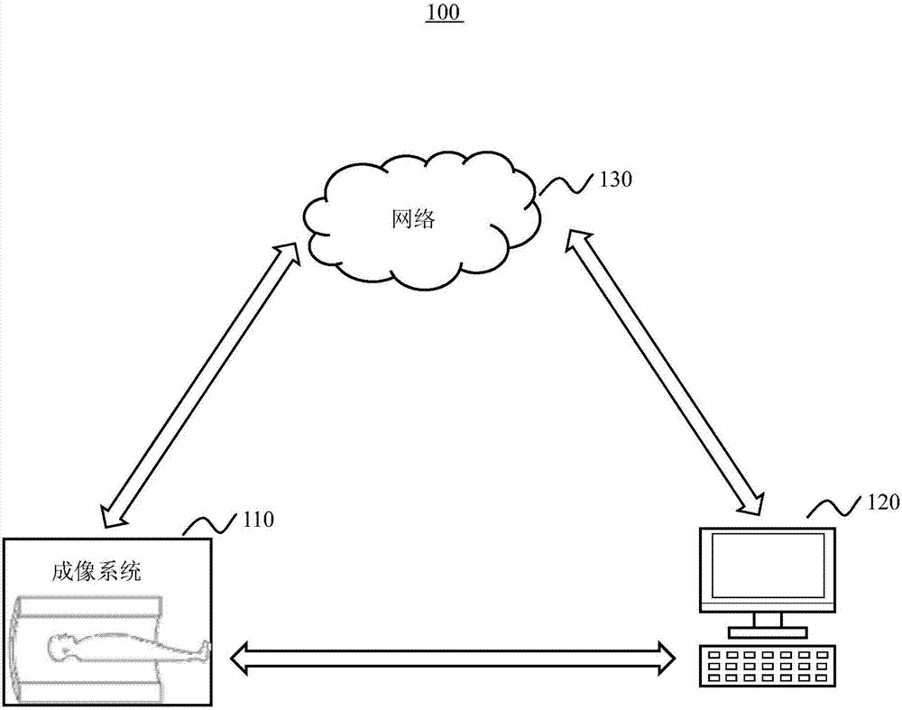
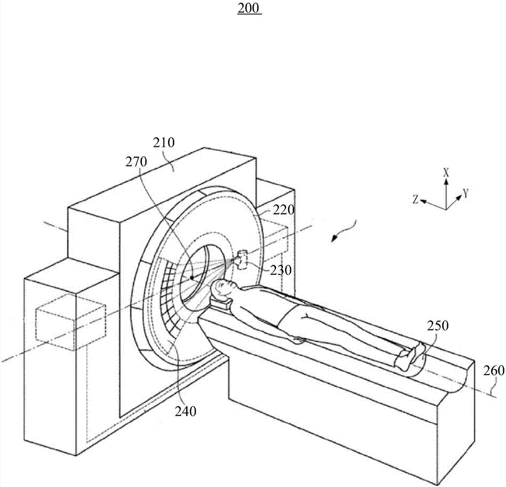
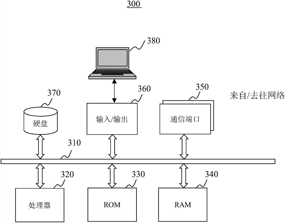
[0032] In order to more clearly illustrate the technical solutions of the embodiments of the present application, the following briefly introduces the drawings that need to be used in the description of the embodiments. Obviously, the accompanying drawings in the following description are only some examples or embodiments of the present application, and those skilled in the art can also apply the present application to other similar scenarios. Unless otherwise apparent from context or otherwise indicated, like reference numerals in the figures represent like structures or operations.

[0033] As indicated in this application and claims, the terms "a", "an", "an" and / or "the" do not refer to the singular and may include the plural unless the context clearly indicates an exception. Generally speaking, the terms "comprising" and "comprising" only suggest the inclusion of clearly identified steps and elements, and these steps and elements do not constitute an exclusive list, and ...


PUM


Abstract

The invention provides an image reconstruction method and device of a medical scanning image. The method includes the following steps: receiving scanning data from medical image equipment; determining projection values of a pair of conjugate rays of each voxel; and performing multi-order interpolation on the projection values of the pair of conjugate rays in a row direction of a detector of the medical scanning image. The reconstruction method of the medical scanning image can improve the longitudinal resolution of a scanned object.

Description

technical field [0001] The present invention mainly relates to medical scanning image processing, in particular to a method and device for reconstructing medical scanning images. Background technique [0002] Computed Tomography (CT) technology is based on the fact that different substances have different attenuation properties for rays. It uses radioactivity to irradiate the measured object from all directions, measures the intensity of the rays passing through the object, and calculates it through a certain reconstruction algorithm. The linear attenuation coefficient of the material at each point inside the object for the rays, so as to obtain the tomographic image of the measured object. Radiological diagnostic technology. [0003] The CT image data obtained by the CT scan is stored after image reconstruction for viewing by an operator. When evaluating the reconstructed image quality of high-resolution CT imaging, the slice sensitivity profile (Slice Sensitivity Profile,...

Claims


Application Information

Patent Timeline
Patent Type & Authority Applications(China)
IPC IPC(8): G06T7/00G06T3/40
CPCG06T3/4007G06T7/0012G06T2207/10081G06T2207/10116G06T2207/30004
Inventor 鲍园王鑫全国涛李翔
Owner SHANGHAI UNITED IMAGING HEALTHCARE
Learn More