End-to-end chest CT image segmentation method based on fully convolutional neural network

A convolutional neural network and CT image technology, applied in the field of medical image processing, can solve the problems of deformation model sensitivity, segmentation result error, and Snake model without standardized segmentation process, so as to achieve the effect of simplifying the process

Inactive Publication Date: 2017-09-26
NANJING UNIV OF POSTS & TELECOMM
View PDF1 Cites 40 Cited by
  • Summary
  • Abstract
  • Description
  • Claims
  • Application Information

AI Technical Summary

Problems solved by technology

However, these segmentation methods also have certain shortcomings. The detection of concave and convex points is more sensitive to the deformation model. The parameters and initialization of each model in the curvature analysis method will have certain errors. The Snake model does not have a standardized segmentation process, and the segmentation results will appear. certain error

Method used


Image

  • End-to-end chest CT image segmentation method based on fully convolutional neural network
  • End-to-end chest CT image segmentation method based on fully convolutional neural network
  • End-to-end chest CT image segmentation method based on fully convolutional neural network

Examples


Embodiment Construction

[0042] The present invention is described in further detail now in conjunction with accompanying drawing. The drawings described here are used to provide a further understanding of the present invention and constitute a part of the application. The schematic embodiments of the present invention and their illustrations are only used to explain the present invention, and do not constitute improper limitations to the present invention.

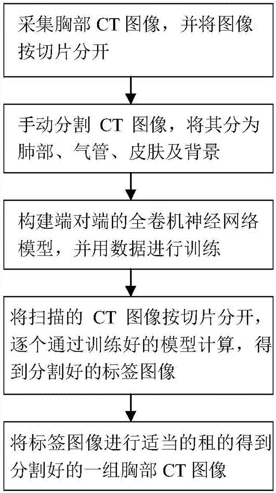
[0043] Such as figure 1 Shown, the present invention comprises the following steps:

[0044] Step 1: Clinically scan k groups of chest CT images, and separate each group of CT images according to each slice as a training sample;

[0045] In the process of obtaining chest CT images, CT scanning is a spiral scan. The scanning process is generally that when the X-ray light source and detector rotate around the patient, the patient's bed moves slowly along the direction of the rotation axis. . Modern CT uses two-dimensional multi-row detectors to ...


PUM


Abstract

The invention discloses an end-to-end chest CT image segmentation method based on a fully convolutional neural network. The method comprises the steps of firstly performing clinic scanning for obtaining k sets of chest CT images, and dividing each set of CT images according to each slice as a training sample; then performing manual cutting on each training sample and a testing sample by professional medical care personnel, dividing the image to four parts, namely a lung part, a tracheae art, a skin part and a background part; then constructing an end-to-end fully convolutional neural network for training marked chest CT training data, and obtaining a trained parameter model; separating the slices of the scanned CT image, and inputting the slices into the trained model one by one, and obtaining segmented output models; and finally combining the output models, thereby obtaining a segmented chest CT image model. The convolutional neural network model which utilizes an image neighborhood content for performing characteristic extraction can perform dense predication on the chest CT image and furthermore simplifies an image segmentation process.

Description

technical field [0001] The invention belongs to the field of medical image processing, and in particular relates to an end-to-end chest CT image segmentation method based on a fully convolutional neural network. Background technique [0002] With the continuous and rapid development of medical imaging and computer technology, the use of computer technology to analyze clinical image data has improved the probability of successful disease prevention and treatment. Computed tomography (CT) is most commonly used in the diagnosis and detection of thoracic diseases. Since CT can provide high-resolution scanning images for various organs or tissues of the chest, it is very important to make full use of CT scanning images for the detection of lung diseases, such as lung cancer, pulmonary nodules and other diseases. In the computer-aided diagnosis system, accurate segmentation of lung CT scan images is the basis and premise of subsequent chest function analysis and three-dimensional...

Claims


Application Information

Patent Timeline
Patent Type & Authority Applications(China)
IPC IPC(8): G06T7/00G06T7/10
CPCG06T7/0012G06T7/10G06T2207/10081G06T2207/20081G06T2207/20084G06T2207/30061
Inventor 冒凯鹏谢世朋
Owner NANJING UNIV OF POSTS & TELECOMM
Why Eureka
Try Eureka