Eye fundus image based three-dimensional model building method and device for retinal vessel

A retinal blood vessel and three-dimensional model technology, which is applied in the field of retinal blood vessel three-dimensional model construction method and its device based on fundus images, and can solve problems such as complex operation

Inactive Publication Date: 2017-09-26
CHONGQING UNIV
View PDF3 Cites 7 Cited by
  • Summary
  • Abstract
  • Description
  • Claims
  • Application Information

AI Technical Summary

Problems solved by technology

[0004] However, the above three types of methods are more complicated to o

Method used


Image

  • Eye fundus image based three-dimensional model building method and device for retinal vessel
  • Eye fundus image based three-dimensional model building method and device for retinal vessel
  • Eye fundus image based three-dimensional model building method and device for retinal vessel

Examples


Embodiment 1

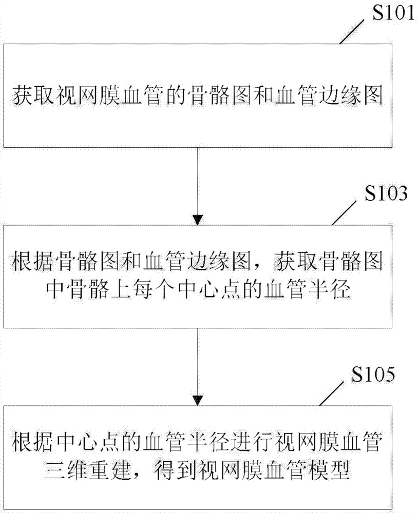
[0065] see figure 1 , is a flow chart of an embodiment of a method for three-dimensional reconstruction of retinal vessels based on fundus images of the present invention. Specifically, in this embodiment, the method for three-dimensional reconstruction of retinal blood vessels includes steps:

[0066] S101. Obtain a bone map corresponding to a skeleton of a retinal vessel and a vessel edge map respectively.

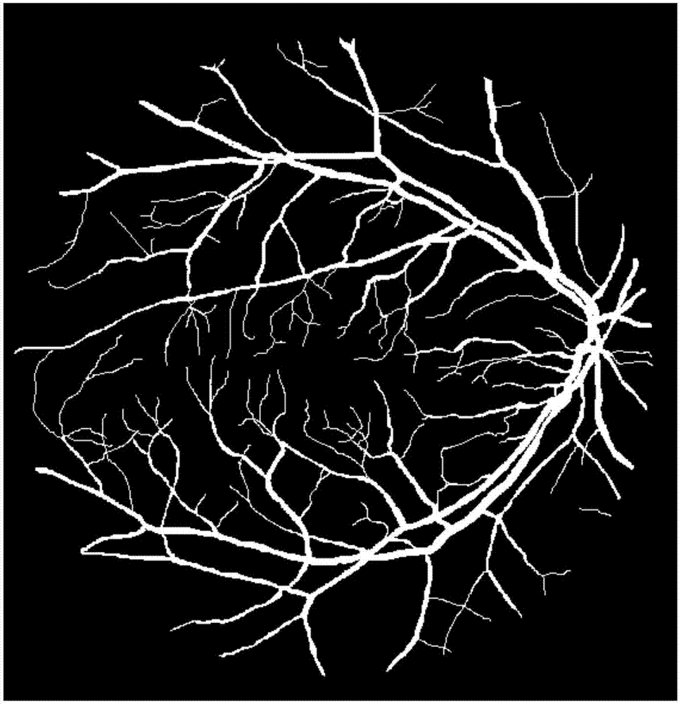
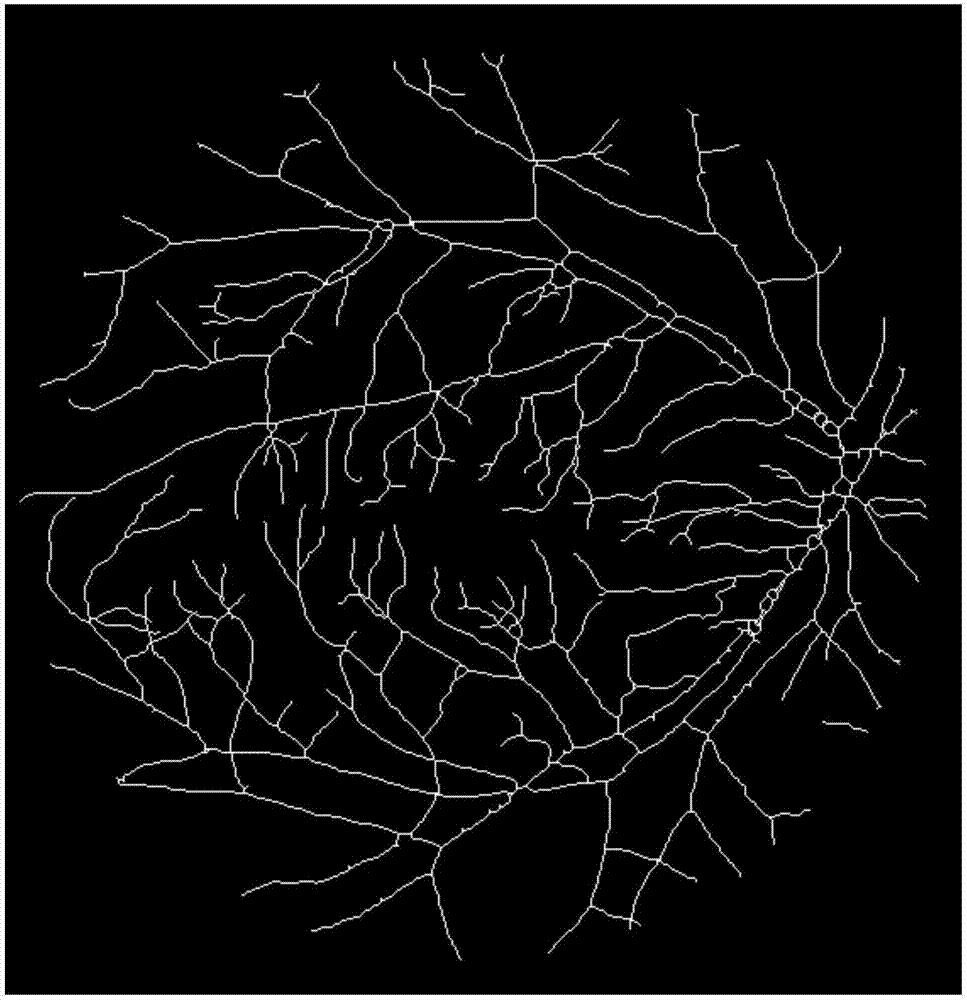
[0067] Since the skeleton is an important topological description of the geometric shape of the image, using the skeleton to represent the original image can reduce the redundant information of the image and highlight its characteristic information while maintaining the important topological features of the image. Therefore, in this embodiment, preprocessing is performed on the original binary image of the retinal blood vessel, so as to obtain its corresponding bone map and blood vessel edge map respectively.

[0068] In a specific embodiment, by calling the bwmorph fu...

Embodiment 2

[0091] Although each center point on the bone is used as the center of the sphere, and the corresponding vascular radius is formed as a small sphere, and then all spheres are combined to obtain a union to obtain a three-dimensional model of retinal blood vessels, but if the pixel points on the bone map (ie Center point) is discontinuous, then the blood vessels in the three-dimensional model of retinal blood vessels constructed based on the skeleton diagram will have abnormally raised parts, such as Figure 5a shown. One of the reasons for this result is: as shown in the figure, when returning the scan radius value in the 45° oblique direction (that is, the preset initial value of the blood vessel radius or the adjusted initial value of the blood vessel radius), what is returned is The number of pixels between the edge of the blood vessel and the center point, but what should actually be returned is the radius value in this direction multiplied by go back. Based on this, the...

Embodiment 3

[0123] The present invention also provides a method for reconstructing a three-dimensional model of a retinal vessel based on a fundus image, which will be described in detail below with reference to specific embodiments and accompanying drawings.

[0124] Since the ball is drawn according to each center point in the skeleton diagram and the corresponding optimized vessel radius, and then the union is obtained to obtain the three-dimensional model of retinal vessels, that is to say, it is actually composed of a two-dimensional (XY) plane space (skeletal diagram and / or blood vessel edge map) into a three-dimensional space (that is, a three-dimensional model, XYZ), then two consecutive points may become non-adjacent and discontinuous because of the large difference in Z values ​​in the third dimension, as shown in the following table three shown.

[0125] Table 3 Coordinate data of the center point before the difference

[0126]

[0127] To make the continuous points in the ...


PUM


Abstract

The invention discloses an eye fundus image based three-dimensional model building method for a retinal vessel. According to the method, a skeleton image and a vessel edge image of the retinal vessel are obtained respectively, then a vessel radius corresponding to each central point on a skeleton is calculated according to the skeleton image and the vessel edge image, then balls are drawn with the central points as ball centers and the vessel radiuses as the radiuses, union is solved, and a three-dimensional module of the retinal vessel is obtained. In order to ensure that a three-dimensional model is closer to a real vessel, the vessel radius obtained through calculation is optimized, that is, the three-dimensional model is built according to the optimized vessel radius. Correspondingly, the invention further provides an eye fundus image based three-dimensional model building device for the retinal vessel.

Description

technical field [0001] The invention belongs to the technical field of medical image processing, and in particular relates to a method for building a three-dimensional model of retinal blood vessels based on a fundus image and a device thereof. Background technique [0002] The quantitative assessment of retinal blood vessels can provide useful clinical auxiliary information for the diagnosis of various diseases. Therefore, the research of retinal blood vessels has great significance in clinical medicine. The results of traditional retinal blood vessel detection are two-dimensional images, and it takes a lot of clinical experience for the diagnostician to get more accurate results from the two-dimensional image. The demands are very high. [0003] With the mature development of 3D reconstruction technology, the medical application of 3D reconstruction technology has also become a hot spot. Because the three-dimensional reconstruction of retinal blood vessels can provide do...

Claims


Application Information

Patent Timeline
IPC IPC(8): G06T17/10
CPCG06T17/10G06T2210/41
Inventor 叶志义王贵学唐甜华唐建清聂天宇
Owner CHONGQING UNIV
Why Eureka
Try Eureka