Multi-characteristic fusion monitored retinal blood vessel extraction method

A retinal blood vessel and multi-feature fusion technology, which is applied in the field of supervised retinal blood vessel extraction with multi-feature fusion, can solve the problems of not being able to extract blood vessel information to the maximum extent, micro-vessel easily discrete, and low sensitivity.

Inactive Publication Date: 2017-10-13
JIANGXI UNIV OF SCI & TECH
View PDF2 Cites 29 Cited by
  • Summary
  • Abstract
  • Description
  • Claims
  • Application Information

AI Technical Summary

Problems solved by technology

[0004] The purpose of the present invention is to address the shortcomings of existing retinal vessel segmentation methods, and provide a supervised retinal vessel extraction method with multi-feature fusion, which solves the problem that the microvessels segmented by the existing model are easy to be separated, have low sensitivity, and cannot maximize Extract blood vessel information and other issues

Method used


Image

  • Multi-characteristic fusion monitored retinal blood vessel extraction method
  • Multi-characteristic fusion monitored retinal blood vessel extraction method
  • Multi-characteristic fusion monitored retinal blood vessel extraction method

Examples


Embodiment Construction

[0039] Below in conjunction with specific embodiment, further illustrate the present invention.

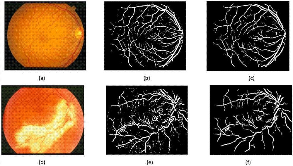
[0040] Explanation of the experiment: the data of the examples involved in the application of the present invention come from the STARE database. The STARE database has 10 fundus images with and without lesions each, and the image size is 605×700 pixels. Secondly, when training the random forest model, 3,000 pixels of blood vessel samples and 7,000 pixels of non-vascular (background) samples are randomly selected from each retinal image, and a total of 200,000 pixels are used as training samples.

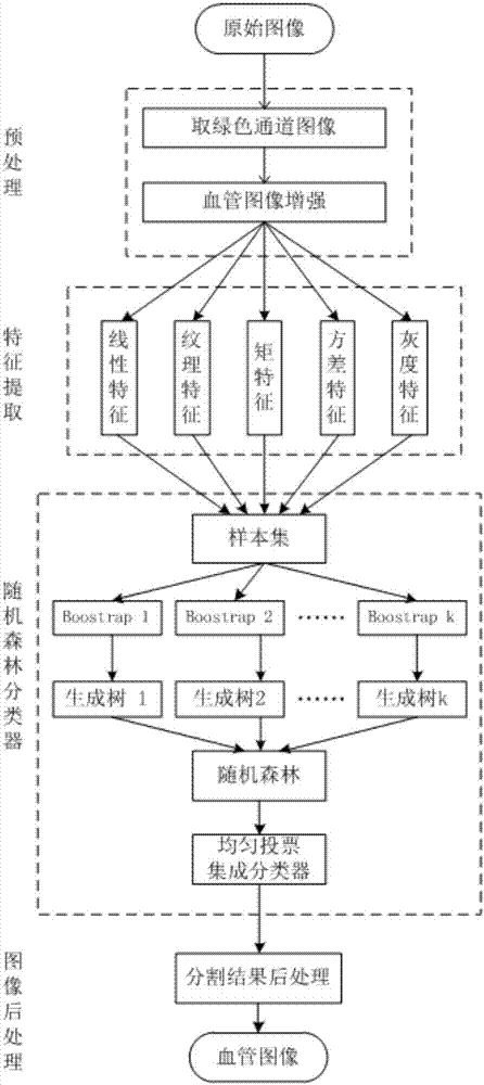
[0041] This embodiment includes four steps: retinal vessel image preprocessing, retinal vessel image feature extraction, random forest classifier training and retinal vessel image post-processing, such as figure 1 shown.

[0042] The specific description is as follows:

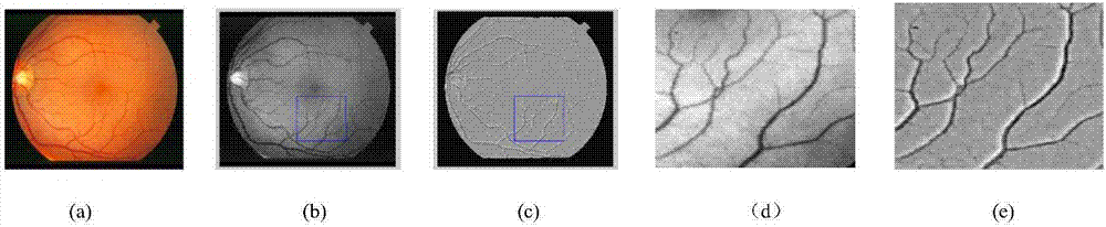
[0043] 1. Retinal blood vessel image preprocessing

[0044] In this embodiment, the green channel retinal image o...


PUM


Abstract

The invention relates to the retinal blood vessel segmentation technology and especially relates to a multi-characteristic fusion monitored retinal blood vessel extraction method. The method comprises steps of step 1, retinal blood vessel image pre-processing; step 2, retinal blood vessel image characteristic extraction; step 3, random forest classifier training; and step 4, retina retinal blood vessel image post-processing. The method is advantaged in that through experiment verification of a DRIVE and STARE eyeground image database, susceptibilities are respectively 0.8354 and 0.8452, accuracies are respectively 94.82% and 95.34%, compared with existing retinal blood vessel segmentation methods in the prior art, the multi-characteristic fusion monitored retinal blood vessel extraction method is integrally better, moreover, disadvantages of the other methods at adjacent blood vessel portions, blood vessel cross sections and capillaries are solved, and segmented blood vessel structures are relatively closer to gold standards and actual blood vessel dimension values.

Description

technical field [0001] The invention relates to retinal vessel segmentation technology, in particular to a supervised retinal vessel extraction method with multi-feature fusion. Background technique [0002] The fundus retinal vascular network has an intricate and multi-layered organizational structure, and many ophthalmic and cardiovascular disease lesions can be directly reflected in the changes in the retinal vascular network structure. Although there is a certain difference between the vascular network and the background, the brightness of the blood vessels gradually changes with the extension of the blood vessels, especially the contrast between the ends of the blood vessels and the background is low, making it difficult to completely segment. Therefore, retinal vessel segmentation technology has always been a hot and difficult point in the field of fundus image analysis. However, information such as the number, branch, angle, and width of retinal blood vessels can be ...

Claims


Application Information

Patent Timeline
Patent Type & Authority Applications(China)
IPC IPC(8): G06T7/11G06T7/44
CPCG06T7/11G06T7/44G06T2207/10004G06T2207/20024G06T2207/20081G06T2207/30041G06T2207/30101
Inventor 梁礼明刘博文黄朝林吴健陈明理
Owner JIANGXI UNIV OF SCI & TECH
Why Eureka
Try Eureka