Unsupervised cervical cell image automatic segmentation method and system

A cervical cell, automatic segmentation technology, applied in the field of medical cell image processing, can solve problems such as unsupervised cervical cell image

Active Publication Date: 2017-10-17
深思考人工智能机器人科技(北京)有限公司
View PDF7 Cites 31 Cited by
  • Summary
  • Abstract
  • Description
  • Claims
  • Application Information

AI Technical Summary

Problems solved by technology

[0005] The purpose of the present invention is to overcome the above-mentioned problems existing in the current cervical cell segmentation, and provide an unsupervised automatic segmentation method for cervical cell images, which can solve the segmentation problem of single independent cells and overlapping cluster cells

Method used


Image

  • Unsupervised cervical cell image automatic segmentation method and system
  • Unsupervised cervical cell image automatic segmentation method and system
  • Unsupervised cervical cell image automatic segmentation method and system

Examples


Embodiment Construction

[0034] The preferred embodiments of the present invention will be described in detail below in conjunction with the accompanying drawings, so that the advantages and features of the present invention can be more easily understood by those skilled in the art, so as to define the protection scope of the present invention more clearly.

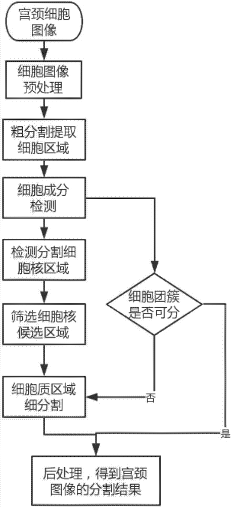
[0035] Such as figure 1 As shown, an unsupervised automatic segmentation method for cervical cell images solves the problems of low accuracy of cervical cell segmentation and effective segmentation of cell clusters. The method includes:

[0036] Step 1) Preprocessing the cervical cell image, the specific process is: using bilateral filtering to denoise, morphological processing and histogram equalization to enhance cell edges and increase contrast;

[0037] For the background clutter of cervical cells, bilateral filtering is used to denoise, and the filter is composed of two functions. A function is determined by the geometric distance of the fi...


PUM


Abstract

The invention discloses an unsupervised cervical cell image automatic segmentation method. The method comprises the following steps of 1) preprocessing a cervical cell image; 2) subjecting the preprocessed cell image to the pre-background rough segmentation, and extracting a region to which cells belong; 3) subjecting the roughly segmented cell image to the detection and segmentation of cell components, and segmenting cells of different types by using a rapid region convolution neural network; 4) detecting and segmenting the cell nucleuses of cervical cells; 5) according to the characteristic parameters of the cell nucleuses, screening the cell nucleuses to obtain final candidate cell nucleuses; 6) judging whether the cell types obtained in the step 3) are multicellular spheroids or not; if not, segmenting a cytoplasmic region by using an activity contour model and a prior template; otherwise, conducting the step 7); 7) conducting the post-treatment based on the segmentation result of cell nucleus and cytoplasm, and the domain knowledge. In this way, the effective segmentation of a whole cervical cell is completed.

Description

technical field [0001] The invention relates to the field of medical cell image processing, in particular to an unsupervised automatic segmentation method and system for cervical cell images. Background technique [0002] Cervical cell artificial intelligence-assisted film reading itself will play a positive role in reducing the labor intensity of film readers, improving the accuracy and work efficiency of film reading, and being applied to large-scale cervical cancer screening. It follows the medical market. The development law and needs are in line with the research layout of national health and medical big data. Accurately locating the cytoplasm and nucleus of each cell from the cervical cell image, and segmenting the two from the entire image is an important basis for the realization of artificial intelligence-assisted film reading of cervical cells. [0003] How to segment cervical cells is a problem worth exploring. Cell images often have problems of overlapping cells...

Claims


Application Information

Patent Timeline
Patent Type & Authority Applications(China)
IPC IPC(8): G06T7/194G06N3/04G06T7/11
CPCG06N3/04G06T7/11G06T7/194G06T2207/30004
Inventor 杨志明李亚伟
Owner 深思考人工智能机器人科技(北京)有限公司
Why Eureka
Try Eureka