
Breast tumor classification method based on differentiated convolutional neural network and breast tumor classification device based on differentiated convolutional neural network

A convolutional neural network and breast tumor technology, applied in the field of breast tumor classification and device based on discriminative convolutional neural network, can solve the problem of poor generalization performance of manual design, difficult to obtain effective information of classification performance, and difficult to feature Learning and other problems to achieve the effect of improving tumor classification performance, avoiding artificial design features, and enhancing discrimination

Active Publication Date: 2018-03-02
SHANDONG UNIV OF FINANCE & ECONOMICS
View PDF2 Cites 26 Cited by
  • Summary
  • Abstract
  • Description
  • Claims
  • Application Information

AI Technical Summary

Problems solved by technology

[0006] However, existing feature extraction and classification methods have certain limitations
In terms of feature extraction, although texture features are an important clinical distinguishing feature of benign and malignant tumors, there are still some unknown image features that can be used to classify tumors. Satisfactory classification performance
In addition, the generalization performance of artificially designed features is poor, and images (different data) for different devices need to be redesigned
In terms of classification, most of the existing classifiers are shallow models, and it is difficult to fully learn the effective information of the features.

Method used


Image

  • Breast tumor classification method based on differentiated convolutional neural network and breast tumor classification device based on differentiated convolutional neural network
  • Breast tumor classification method based on differentiated convolutional neural network and breast tumor classification device based on differentiated convolutional neural network
  • Breast tumor classification method based on differentiated convolutional neural network and breast tumor classification device based on differentiated convolutional neural network

Examples


Embodiment 1

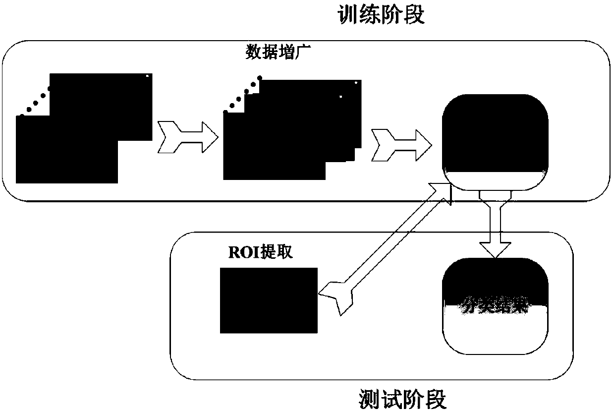
[0035] This embodiment discloses a breast tumor classification method based on a discriminative convolutional neural network, which is divided into two stages of training and testing:

[0036] Training phase:

[0037] Step (11): Using the C-V active contour model to segment the tumor in the ultrasound image, obtain a region of interest (ROI), and select a part as a training image;

[0038] Step (12): Carry out data augmentation to training image, obtain new training set;

[0039] Step (13): constructing a discriminative convolutional neural network model, and calculating model parameters of the discriminative convolutional neural network based on the training set.

[0040] Testing phase:

[0041] Step (14): Obtain a breast ultrasound image to be classified, use the C-V active contour model to segment the tumor in the ultrasound image, and obtain a region of interest (ROI);

[0042] Step (15): Input the ROI into the trained discriminative convolutional neural network to obta...

Embodiment 2

[0061] The purpose of this embodiment is to provide a computing device.

[0062] A breast tumor classification device based on a discriminative convolutional neural network, comprising a memory, a processor, and a computer program stored on the memory and operable on the processor, and the processor implements the following steps when executing the program, including :

[0063] Receive multiple ultrasound images, segment the tumors in them, and obtain training images;

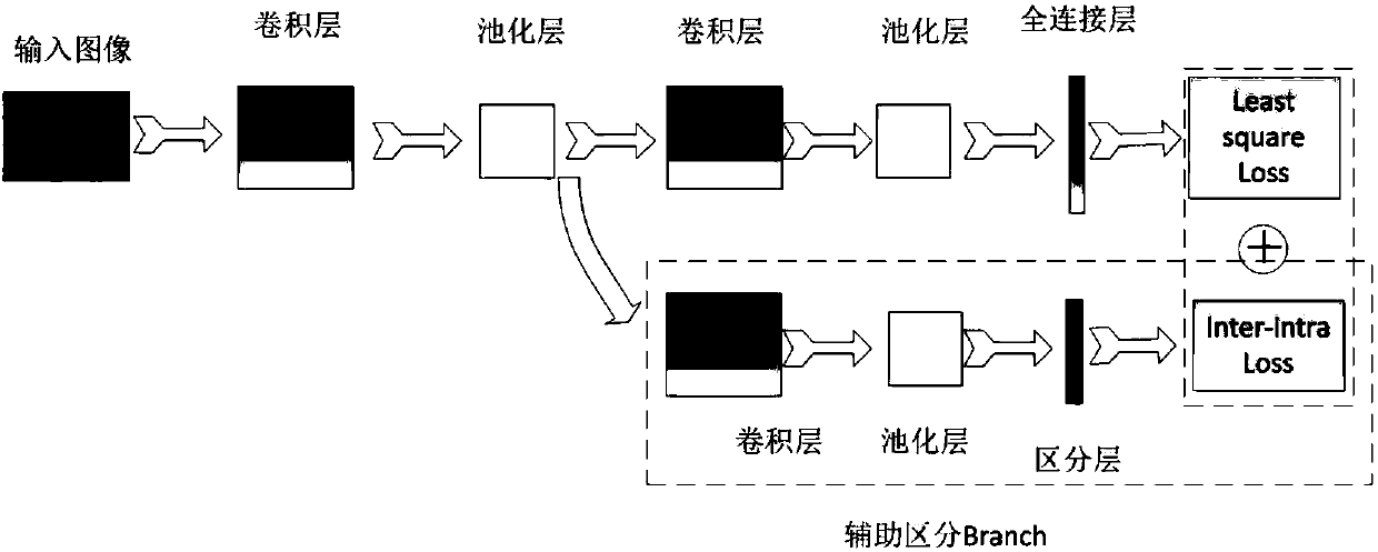
[0064] Build a discriminative convolutional neural network model, calculate the model parameters of the discriminative convolutional neural network based on the training image; wherein, the structure of the discriminative convolutional neural network model is: on the basis of the convolutional neural network Add a discriminative auxiliary branch to access the convolutional layer, pooling layer and fully connected layer;

[0065] receiving a breast ultrasound image to be classified, segmenting the ultrasound i...

Embodiment 3

[0068] The purpose of this embodiment is to provide a computer-readable storage medium.

[0069] A computer-readable storage medium, on which a computer program is stored, and when the program is executed by a processor, the following steps are performed:

[0070] Receive multiple ultrasound images, segment the tumors in them, and obtain training images;

[0071] Build a discriminative convolutional neural network model, calculate the model parameters of the discriminative convolutional neural network based on the training image; wherein, the structure of the discriminative convolutional neural network model is: on the basis of the convolutional neural network Add a discriminative auxiliary branch to access the convolutional layer, pooling layer and fully connected layer;

[0072] receiving a breast ultrasound image to be classified, segmenting the ultrasound image, and obtaining a region of interest;

[0073] The region of interest is input to the discriminative convolution...


PUM


Abstract

The invention discloses a breast tumor classification method based on a differentiated convolutional neural network and a breast tumor classification device based on a differentiated convolutional neural network. The method comprises the steps that the tumor in multiple ultrasonic images is segmented to acquire an area of interest, and data augmentation is performed so that a training set is obtained; a differentiated convolutional neural network model is constructed, and the model parameters of the differentiated convolutional neural network are calculated based on training images, wherein the structure of the differentiated convolutional neural network model is that differentiated auxiliary branches are additionally arranged on the basis of the convolutional neural network, a convolutional layer, a pooling layer and a full connection layer are accessed, and an Inter-intra Loss function is introduced for increasing the similarity between the same classes and the differentiation between different classes; a breast ultrasonic image to be classified is acquired, the ultrasonic image is segmented and the area of interest is acquired; and the area of interest is inputted to the differentiated convolutional neural network so as to obtain the classification result. According to the classification method, the tumor classification performance in the breast ultrasonic image can be effectively enhanced.

Description

technical field [0001] The invention belongs to the field of data classification for medical images, and in particular relates to a breast tumor classification method and device based on a discriminative convolutional neural network. Background technique [0002] For women, breast cancer is one of the diseases with higher morbidity and mortality. Early detection and early treatment is the key to improving treatment efficiency. Medical imaging has become the main way to assist in clinical diagnosis of diseases. Compared with other images such as mammography and nuclear magnetic resonance, ultrasound has the advantages of less radiation, low price, and sensitivity to dense tissue detection. Therefore, ultrasound images have become one of the main tools to assist in the early diagnosis of breast cancer. [0003] Due to the different experience of radiologists, manual diagnosis of breast ultrasound images has a certain degree of subjectivity. The use of computer-aided diagno...

Claims


Application Information

Patent Timeline
IPC IPC(8): G06K9/62G06K9/32G06K9/34G06N3/04
CPCG06V10/25G06V10/267G06V2201/032G06N3/045G06F18/24G06F18/214
Inventor 袭肖明尹义龙孟宪静聂秀山杨璐
Owner SHANDONG UNIV OF FINANCE & ECONOMICS
Learn More