Method for automatically evaluating picture quality of digital pathological scanning image

A technology of digital pathology and scanned images, applied in the field of medical image detection, can solve the problems of wasting time, missing images, unqualified images, etc., and achieve the effect of improving work efficiency, reducing the proportion of non-blurred images, and reducing the time for image quality evaluation

Inactive Publication Date: 2018-04-20
KONFOONG BIOTECH INT
View PDF8 Cites 11 Cited by
  • Summary
  • Abstract
  • Description
  • Claims
  • Application Information

AI Technical Summary

Problems solved by technology

However, the evaluation method of this method is relatively radical. If a single line image of the image is unqualified, the entire image is unqualified, and it is easy to miss some images, resulting in the need to re-scan the pathological slice, which wastes time.
[0009] Therefore, there is an urgent need for a method that can reduce human evaluation, save time, and improve the automatic evaluation of digital pathology image quality, but there is no report on this method at present

Method used


Image

  • Method for automatically evaluating picture quality of digital pathological scanning image
  • Method for automatically evaluating picture quality of digital pathological scanning image
  • Method for automatically evaluating picture quality of digital pathological scanning image

Examples


Embodiment 1

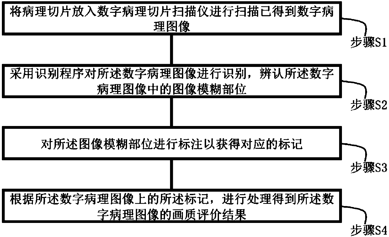
[0050] Such as figure 1 As shown, a method for automatically evaluating the image quality of digital pathological scanning images includes a pathological slide scanner, and the pathological slide scanner has a built-in recognition program, including the following steps:

[0051] Step S1, putting the pathological slice into a digital pathological slice scanner to scan to obtain a digital pathological image;

[0052] Step S2, using a recognition program to recognize the digital pathological image, and identify blurred parts in the digital pathological image;

[0053] Step S3, marking the blurred part of the image to obtain a corresponding mark;

[0054] Step S4, performing processing according to the marks on the digital pathological image to obtain an evaluation result of the image quality of the digital pathological image.

[0055] As a preferred embodiment, in the step S2, the recognition program is used to recognize the digital pathological image using a method based on edge...

Embodiment 2

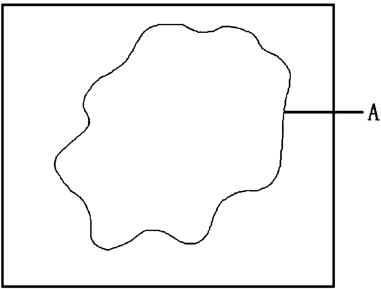
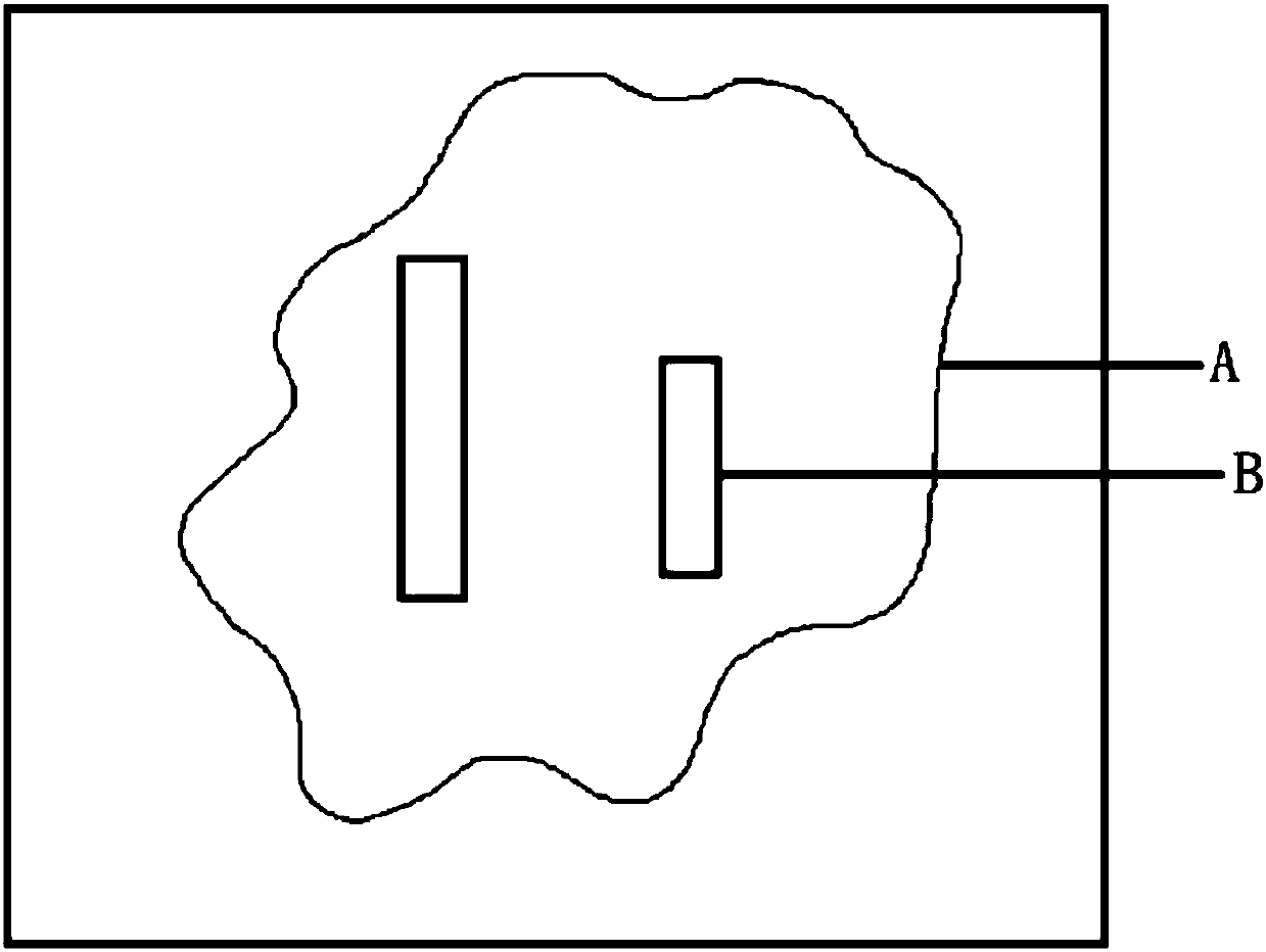
[0094] Such as figure 2 , 3 As shown, a method for automatically evaluating the image quality of digital pathological scanning images of the present invention is applied as follows:

[0095] Step S1, putting the pathological slice into a digital pathological slice scanner to scan to obtain a digital pathological image ( figure 2 shown);

[0096] Step S2, using a recognition program to recognize the digital pathological image, and identify blurred parts in the digital pathological image;

[0097] Step S3, mark the blurred part of the image to obtain the corresponding mark ( image 3 shown);

[0098] Step S4, performing processing according to the marks on the digital pathological image to obtain an evaluation result of the image quality of the digital pathological image.

[0099] The digital image can be evaluated in three different ways: the digital image has 1 cell tissue A, the digital image has 2 markers B in total, and the threshold of the number of markers is not e...


PUM


Abstract

The invention relates to a method for automatically evaluating the picture quality of a digital pathological scanning image. The method comprises a pathological section scanner, wherein an identification program is built in the pathological section scanner. The method comprises the following steps that: S1: putting a pathological section into the pathological section scanner to be scanned to obtain a digital pathological image; S2: adopting an identification program to identify the digital pathological image, and distinguishing an image fuzzy position in the digital pathological image; S3: labeling the image fuzzy position to obtain a corresponding sign; and S4: according to the sign on the digital pathological image, carrying out processing to obtain the picture quality evaluation resultof the digital pathological image. The method has the advantages that the identification program is used for automatically labeling the digital image, and working efficiency is improved; through different evaluation ways, the digital pathological image is effectively screened, and picture quality evaluation time can be effectively shortened.

Description

technical field [0001] The invention relates to the field of medical image detection, in particular to a method for automatically evaluating the image quality of digital pathological scanning images. Background technique [0002] Digital pathology refers to the application of computers and networks in the field of pathology. It is a technology that combines modern digital systems with traditional optical amplification devices. It scans and collects high-resolution digital images through a fully automatic microscope or an optical magnification system, and then uses a computer to automatically stitch and process the obtained images with high precision and multiple fields of view seamlessly to obtain high-quality visual data for use in various fields of pathology. field. [0003] The core of digital pathology is a digital pathological slide scanner that converts pathological slices into digital slices, and the image quality (referred to as image quality) of digital images scan...

Claims


Application Information

Patent Timeline
Patent Type & Authority Applications(China)
IPC IPC(8): G06T7/00G06T7/11G06T7/62
CPCG06T7/0012G06T2207/30168G06T7/11G06T7/62
Inventor 刘炳宪谢菊元王焱辉王克惠张晨晨
Owner KONFOONG BIOTECH INT
Why Eureka
Try Eureka