A preparation method of human airway and cervical vertebra 3D printing model

A 3D printing and cervical spine technology, applied in the field of 3D printing, can solve the problems of complex airway structure and inability to understand the structure, and achieve the effects of reduced risk, low price and cost reduction

Active Publication Date: 2019-10-18
PEKING UNIV THIRD HOSPITAL
View PDF4 Cites 0 Cited by
  • Summary
  • Abstract
  • Description
  • Claims
  • Application Information

AI Technical Summary

Problems solved by technology

[0003] The purpose of the present invention is to provide a method for preparing a 3D printed model of the human airway and cervical spine, to solve the problem that the structure of the difficult airway is complex and there is no way to understand the detailed structure

Method used


Image

  • A preparation method of human airway and cervical vertebra 3D printing model
  • A preparation method of human airway and cervical vertebra 3D printing model
  • A preparation method of human airway and cervical vertebra 3D printing model

Examples


Embodiment Construction

[0035] The present invention will be further described in detail below in conjunction with the accompanying drawings and specific embodiments. There is no software innovation in the present invention.

[0036] The invention provides a method for preparing a human airway and cervical vertebra 3D printing model, comprising the following steps:

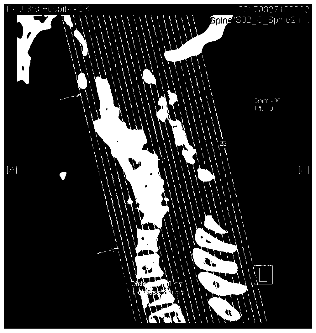
[0037] Step 1. Following the principles of medical ethics, obtain the images and data obtained from the original CT tomography of the patient's intended printing site;

[0038] The image is in Dicom format or JPG format, and the tomogram format and image size must be uniform;

[0039] CT is medical data information, and what 3D printing needs is a closed solid model, so the image is converted through Mimics software.

[0040] Step 2. Carry out three-dimensional reconstruction according to the obtained image and data of the part to be printed;

[0041] What 3D printing needs is a closed solid model, so we need to convert the scanned im...


PUM


Abstract

The invention provides a method for preparing a 3D printed model of a human airway and cervical vertebra. The method comprises the following steps: subjecting a 3D printed hollow airway to 3D solid scanning so as to obtain a 3D scanned image and 3D image data, comparing the obtained 3D scanned image and 3D image data with original CT scanning data, and evaluating the accuracy of the printed hollowairway; and then assembling a printed cervical vertebra and adjacent appendicular bony tissue thereof with the hollow airway. The method provided by the invention can completely reproduce the airway,the cervical vertebra and adjacent anatomical structures, facilitates preoperative simulation of endotracheal intubation in patients with difficult airways, and formulates detailed airway managementstrategies; a hard material is used for printing to print bony structures, and a soft material is used for printing the airway, so the body structure of human beings is authentically reproduced; and to highlight the airway, the airway printing material can be of different colors to facilitate clinical teaching.

Description

technical field [0001] The invention relates to the field of 3D printing, in particular to a method for preparing a 3D printing model of a human airway and cervical spine. Background technique [0002] Cervical degenerative diseases represented by esophageal cervical spondylosis, etc., may be caused by intervertebral disc degeneration secondary to tearing of the anterior longitudinal ligament and subperiosteum, hemorrhage, organization, calcification, and bone spur formation, which will affect the structure of the airway and make the airway The structure of the trachea is more complicated, and in some patients with severe cervical diseases, even experienced anesthesiologists may still find it difficult to successfully perform endotracheal intubation, which is clinically called difficult airway. Without knowing the specific structure of the airway, anesthesiologists cannot successfully perform endotracheal intubation before general anesthesia, and blind intubation will cause ...

Claims


Application Information

Patent Timeline
Patent Type & Authority Patents(China)
IPC IPC(8): B29C64/124B29C64/393B33Y10/00B33Y50/02
Inventor 王军韩彬李正迁刘亚杰张小青
Owner PEKING UNIV THIRD HOSPITAL
Why Eureka
Try Eureka