Automatic analysis method for cone beam CT image three-dimensional craniofacial structure

A CT image, automatic analysis technology, applied in the field of computer vision and oral clinical medicine, can solve the problems of processing fine structure, high online computing cost, etc.

Active Publication Date: 2018-06-26
PEKING UNIV
View PDF8 Cites 17 Cited by
  • Summary
  • Abstract
  • Description
  • Claims
  • Application Information

AI Technical Summary

Problems solved by technology

However, methods based on statistical surface or grayscale models are difficult to deal with fine structures in the reduced subspace
Annotation migration and fusion using image registration or template deformation methods often have high online computing costs
In ...

Method used


Image

  • Automatic analysis method for cone beam CT image three-dimensional craniofacial structure
  • Automatic analysis method for cone beam CT image three-dimensional craniofacial structure
  • Automatic analysis method for cone beam CT image three-dimensional craniofacial structure

Examples


Embodiment Construction

[0038] In the following, the present invention is further described through embodiments with reference to the drawings, but the scope of the present invention is not limited in any way.

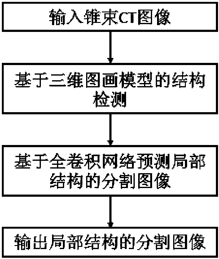
[0039] The present invention provides an automatic analysis method for three-dimensional craniofacial structure of cone-beam CT images. Based on a pictorial model and a fully convolutional neural network, it can realize interest in cone-beam CT images from the input cone-beam CT images. The 3D craniofacial structure is automatically segmented and annotated to obtain automatic analysis and segmentation of stable structures.

[0040] The present invention automatically detects and annotates the jaw bone, zygomatic arch, and anterior skull base in the cone-beam CT image, in which the picture model is used to detect the anatomical structure of interest on the three-dimensional cone-beam CT image and obtain the three-dimensional image where the structure is located. Piece. A fully convolutional neural...


PUM


Abstract

The invention discloses an automatic analysis method for the cone beam CT image three-dimensional craniofacial structure. Based on a picture model and the full convolutional neural network, the picture model is trained for automatic detection and positioning of an anatomical structure, and the full convolutional neural network is utilized to construct mapping between a cone beam CT image of the anatomical structure and a corresponding marked image. At the test stage, for an input three-dimensional cone beam CT image, firstly, the picture model is utilized to detect the spatial position of theanatomical structure of interests, secondly, the full convolutional neural network is utilized to estimate automatic marking of image subblocks of the structure, and automatic three-dimensional craniofacial structure analysis is realized. The method is advantaged in that the three-dimensional craniofacial structure of interests in the cone beam CT image can be automatically segmented and marked, automatic analysis and segmentation of the stable structure are acquired, and the method can be applied to evaluation of marking and the curative effect of an oral orthodontic treatment plan.

Description

Technical field [0001] The invention relates to the field of computer vision and oral clinical medicine, in particular to a method for automatically analyzing three-dimensional craniofacial structures in cone beam CT images. Background technique [0002] The automatic analysis of craniofacial structure is the basis for clinical orthodontic treatment evaluation and surgical prediction. For example, overlapping cone-beam CT images before and after treatment to visualize and evaluate changes in craniofacial morphology caused by treatment and growth depends on cone-beam CT The locally stable structure of the segmentation in the image. Taking into account the local fine bone structure, it is still difficult to automatically segment the relatively stable structures in craniofacial orthodontics, such as the zygomatic arch and anterior skull base. Manual interactive segmentation is time-consuming and depends on the experience of relevant personnel. Cone-beam CT images have a relatively...

Claims


Application Information

Patent Timeline
IPC IPC(8): G06T7/11G06T7/33
CPCG06T2207/10012G06T2207/10081G06T2207/30016
Inventor 裴玉茹秦海芳易芸皑郭玉珂马赓宇许天民查红彬
Owner PEKING UNIV
Why Eureka
Try Eureka