A Segmentation Method of Left and Right Hemibrain

A technology of left and right and cerebrospinal, applied in the field of image processing, can solve problems such as affecting the stability of fitting results, and achieve the effect of reducing radiation-induced brain damage and improving convenience

Active Publication Date: 2020-06-02
SHANGHAI UNITED IMAGING HEALTHCARE
View PDF8 Cites 0 Cited by
  • Summary
  • Abstract
  • Description
  • Claims
  • Application Information

AI Technical Summary

Problems solved by technology

Since the falx only exists in about 30% of the CT image level, and the impact of CT image quality or lesions, there must be many interference points in the detected feature points of the falx, which will affect the stability of the fitting result

Method used


Image

  • A Segmentation Method of Left and Right Hemibrain
  • A Segmentation Method of Left and Right Hemibrain
  • A Segmentation Method of Left and Right Hemibrain

Examples


Embodiment Construction

[0039] A method for dividing the left and right hemispheres proposed by the present invention will be described in further detail below in conjunction with the accompanying drawings and specific embodiments. Advantages and features of the present invention will be apparent from the following description and claims. It should be noted that all the drawings are in a very simplified form and use imprecise scales, and are only used to facilitate and clearly assist the purpose of illustrating the embodiments of the present invention.

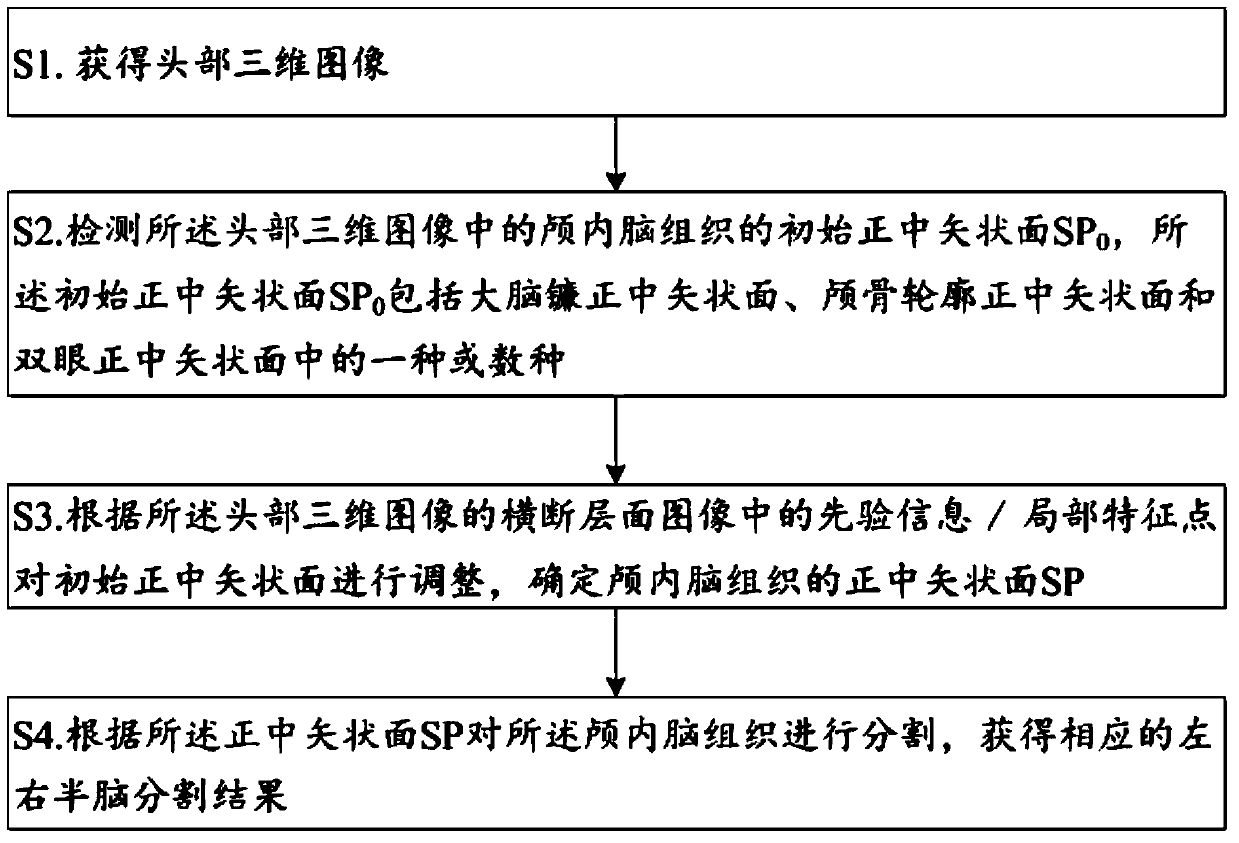
[0040] A kind of segmentation method of left and right hemispheres of the embodiment of the present invention, comprises the following steps:

[0041] S1. Obtain a three-dimensional image of the head; the three-dimensional image of the head is a brain CT image or an MR image;

[0042] S2. Detecting the initial median sagittal plane SP of the intracranial brain tissue in the three-dimensional image of the head 0 , the initial midsagittal plane SP 0...


PUM


Abstract

The invention discloses a segmentation method of the left and right hemibrains, comprising the following steps: S1. Obtaining a three-dimensional image of the head; S2. Detecting the initial median sagittal plane SP of the intracranial brain tissue in the three-dimensional image of the head 0 , the initial midsagittal plane SP 0 Including one or more of the median sagittal plane of the falx cerebrum, the median sagittal plane of the skull outline, and the median sagittal plane of both eyes; S3. According to the prior information / local feature points in the cross-sectional image of the three-dimensional head image Adjusting the initial median sagittal plane to determine the median sagittal plane SP of the intracranial brain tissue; S4. Segmenting the intracranial brain tissue according to the median sagittal plane SP to obtain corresponding left and right hemibrain segmentation results; The method of the invention can quickly and stably divide the left and right hemispheres.

Description

technical field [0001] The invention relates to the technical field of image processing, in particular to a method for dividing the left and right hemibrains used for estimating radiation brain damage. Background technique [0002] Radiation brain injury is a common complication after radiotherapy for extracranial and non-neoplastic lesions. Severe cases greatly affect the quality of life and survival of patients. Solving the contradiction between injury and treatment has always been a difficult and hot topic in research. How to minimize the occurrence of radiation-induced brain injury is a clinical problem that needs to be solved urgently. [0003] At this stage, in the design stage of radiotherapy plan, the strategy of using the results of left and right hemisphere segmentation to predict radiation brain injury is relatively new and unique. Compared with MR images, soft tissue contrast is poor in CT images, and therefore, relatively little is known about the segmentation...

Claims


Application Information

Patent Timeline
Patent Type & Authority Patents(China)
IPC IPC(8): G06T7/11G06T7/136G06T7/168G06K9/32
CPCG06T2207/30008G06T2207/30016G06T2207/10088G06T2207/10081G06V10/242G06V10/25
Inventor 韩妙飞马金凤李强
Owner SHANGHAI UNITED IMAGING HEALTHCARE
Why Eureka
Try Eureka