

Lung CT image data segmentation method and system
A data segmentation and image data technology, applied in the fields of medical image processing and artificial intelligence, can solve the problems of low efficiency, long time consumption and slow speed of lung CT image data, and achieve the effect of improving processing efficiency and facilitating intuitive display.
- Summary
- Abstract
- Description
- Claims
- Application Information
AI Technical Summary
Problems solved by technology
Method used
Image

Examples
Embodiment Construction
[0053] The technical solutions of the present invention will be further described below in conjunction with the accompanying drawings and embodiments.
[0054] see figure 1 , the embodiment of the present invention takes the lung CT image data of patients with new coronary pneumonia as an example to illustrate the lung CT image data segmentation method, including the following steps:
[0055] Step S101. Obtain CT plain scan image data of the lungs of patients with COVID-19.
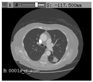
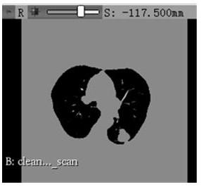
[0056] The subjects of lung CT plain scan image data collection of COVID-19 patients come from COVID-19 patients of different ages in multiple authorized medical centers and regions. During the data collection process, the personal information of patients with new coronary pneumonia should be kept strictly confidential to prevent the leakage of personal information during the collection process. E.g, Figure 3a It is a schematic diagram of the original lung CT plain scan image of a patient with new coron...
PUM
 Abstract
Description
Claims
Application Information