
Optical system of portable fundus camera

An optical system and portable technology, applied in the fields of ophthalmoscope, medical science, eye testing equipment, etc., can solve the problems of expensive equipment, equipment maintenance costs and inspection costs, etc., and achieve low production costs, simple structure, and improved imaging. quality effect

Inactive Publication Date: 2018-07-24
TIANJIN POLYTECHNIC UNIV
View PDF15 Cites 4 Cited by
  • Summary
  • Abstract
  • Description
  • Claims
  • Application Information

AI Technical Summary

Problems solved by technology

Large-scale desktop fundus camera equipment is often expensive, and requires professional medical staff to operate, equipment maintenance and inspection costs are expensive

Method used


Image

  • Optical system of portable fundus camera
  • Optical system of portable fundus camera
  • Optical system of portable fundus camera

Examples


Embodiment Construction

[0029] The present invention provides a portable fundus camera optical system. In order to make the purpose, technical solution and effect of the present invention clearer and clearer, the present invention will be further described in detail below with reference to the accompanying drawings and examples.

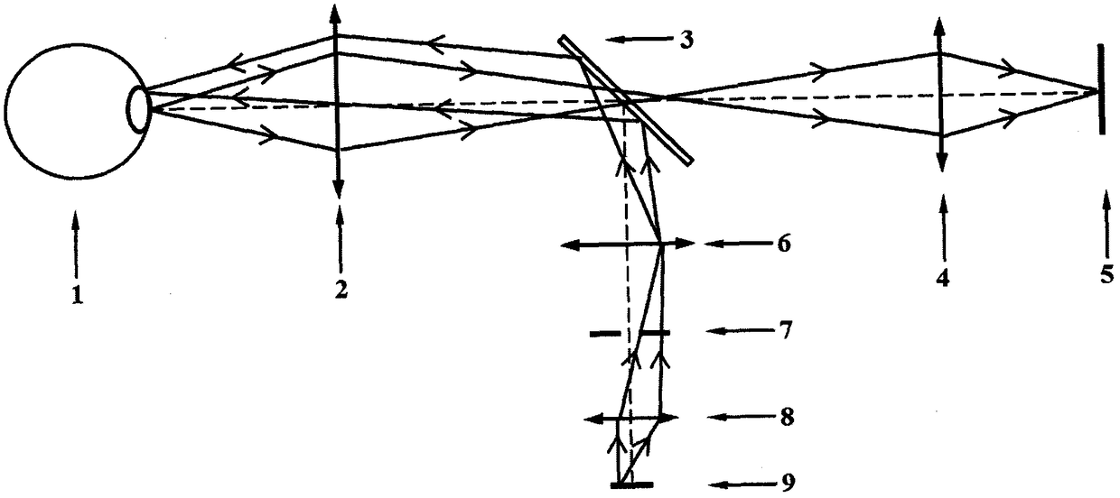
[0030] The structural form of the embodiment of the present invention is designed for the 3 million pixel, 2 / 3 inch CCD existing in the market as a receiving device, the full field of view is 30°, and the total system length does not exceed 250mm. Since the pupil limits the width of the beam entering the human eye and also limits the width of the beam exiting the human eye, it is very appropriate to use the position of the pupil as the position of the aperture stop of the imaging system. Under normal circumstances, the normal adjustment range of the pupil of the human eye is generally 3mm to 7mm, so the entrance pupil diameter of the system can be taken as 4mm. In addition,...


PUM

PropertyMeasurementUnit
Diameteraaaaaaaaaa
Outer diameteraaaaaaaaaa
Wavelengthaaaaaaaaaa

Abstract

The invention belongs to the technical field of fundus medical science, concretely relating to an optical system of a portable fundus camera. Fundus images of human eyes are acquired by using an optical imaging method in order to determine pathologies and lesions of fundus diseases. The optical system of the portable fundus camera comprises two parts including an imaging system and an illuminatingsystem. The imaging system comprises a tested human eye, an ocular objective lens, an imaging objective lens and a reception device. The illuminating system comprises the tested eye, the ocular objective lens, a half-reflecting pellicle mirror, an illuminating objective lens, an annular diaphragm, a collecting mirror and a light source. The included angle formed between the half-reflecting pellicle mirror and a primary optical axis is 45 degrees. A dark light source commonly used in medical science is adopted by the light source. The reception device employs CCD or CMOS. Optical elements adopted by the system are all ordinary spherical mirrors which are convenient to process and manufacture. In this sense, cost is reduced. In addition, the system has just a few optical elements so that the structure is simple. The system is convenient to operate.

Description

technical field [0001] The invention relates to an ophthalmic optical instrument, in particular to a portable fundus camera optical system, which belongs to the field of medical fundus imaging. Background technique [0002] The human retina, also known as the fundus, is an important part of the human eye. The retina is a thin film with a highly complex structure, and a large number of capillaries are distributed on it. It is the only deep microvascular network in the human body that can be directly observed without trauma. Observing the vascular veins on the retina of the human eye can provide an important basis for doctors to diagnose various eye diseases and even systemic diseases. For example, the incidence of hypertension, hyperlipidemia, kidney disease, diabetes, pregnancy-induced hypertension, coronary heart disease and other diseases in our country is increasing year by year, and the early stage of the disease will be reflected in the fundus. As far as diabetes is c...

Claims


Application Information

Patent Timeline
IPC IPC(8): A61B3/12A61B3/14
CPCA61B3/0008A61B3/0016A61B3/12A61B3/14A61B2560/0431
Inventor 肖志涛耿磊王梦蝶张芳吴骏王雯
Owner TIANJIN POLYTECHNIC UNIV
Learn More