
Magnet for treatment under endoscope

A technology using magnets and magnets, which is applied in the field of magnets for endoscopic treatment, can solve the problems of long recovery time for oral feeding, achieve the effects of short recovery time for oral feeding, convenient operation, and short hospitalization period

Active Publication Date: 2018-07-24
WEST CHINA HOSPITAL SICHUAN UNIV
View PDF9 Cites 0 Cited by
  • Summary
  • Abstract
  • Description
  • Claims
  • Application Information

AI Technical Summary

Problems solved by technology

However, the above method takes a long time to recover oral food

Method used


Image

  • Magnet for treatment under endoscope
  • Magnet for treatment under endoscope
  • Magnet for treatment under endoscope

Examples


Embodiment 1

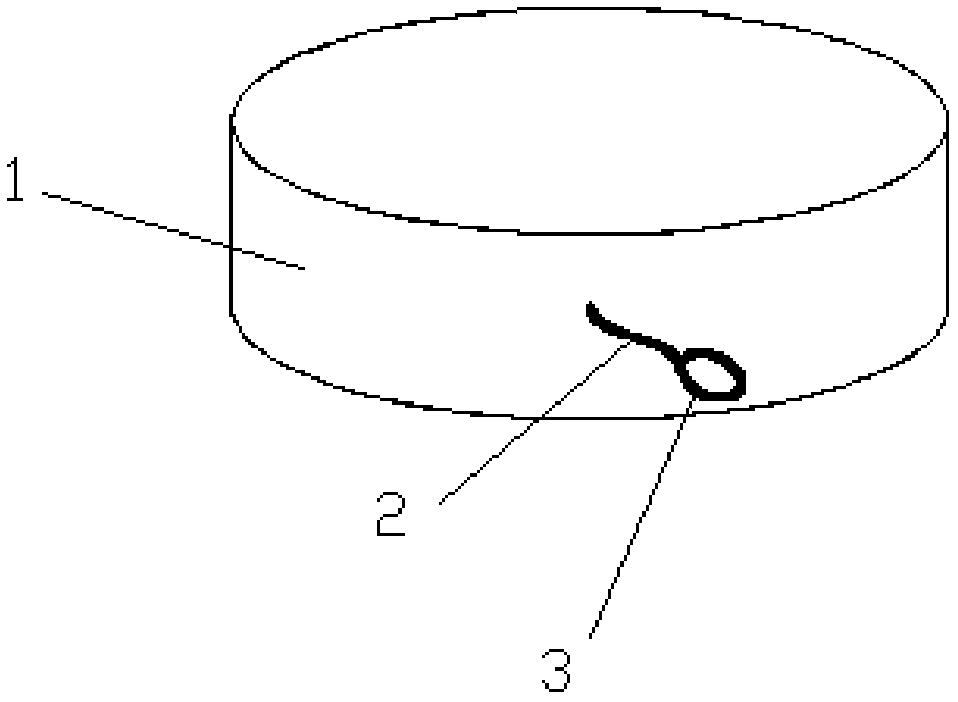
[0028] Such as figure 1 As shown, the magnet for endoscopic treatment disclosed in this embodiment includes at least one magnet body 1, the magnetic poles of the magnet body 1 are at its upper and lower ends, the magnet body 1 is provided with at least one connecting rope 2, and one end of the connecting rope 2 Connected with the magnet body 1, the other end of the connecting rope 2 is a rope ring 3, and the rope ring 3 is used for the biopsy forceps to extend into the jaws, so as to clamp the magnet body 1 conveniently. The connecting rope 2 and the rope ring 3 are integrally manufactured.

[0029] Further, the end faces of the upper and lower ends of the magnet body 1 are planes parallel to each other. The cross section of the magnet body 1 is circular, and the diameter of the cross section of the magnet body 1 is greater than the height of the magnet body 1 . In this embodiment, the magnet body 1 is a solid structure; the connecting rope 2 is fixedly connected to the side...

Embodiment 2

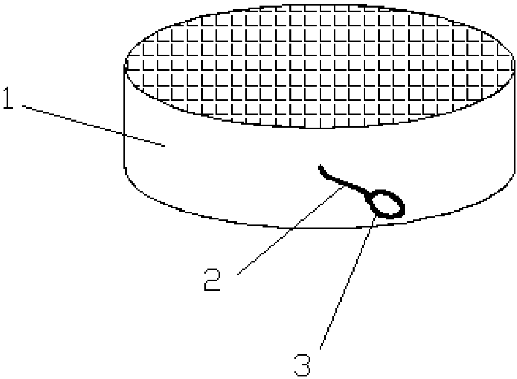
[0032] The difference between this embodiment and embodiment 1 is: as figure 2 As shown, the end surfaces of the upper and lower ends of the magnet body 1 are rough surfaces. The rough surface can be a toothed surface or several protrusions, or knurled. Rough surface can accelerate the rate of ischemic necrosis of the junctional mucosa.

Embodiment 3

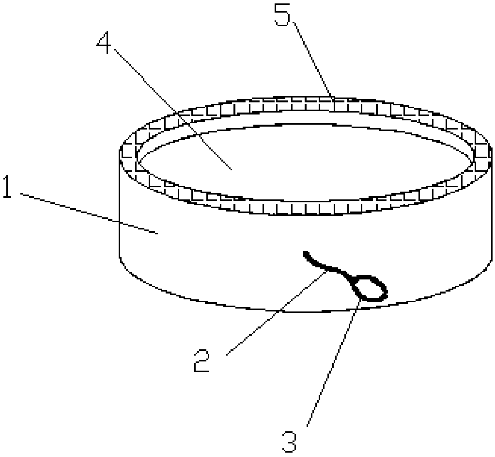
[0034] The difference between this embodiment and embodiment 2 is: as image 3 As shown, the center of both ends of the magnet body 1 is sunken inward to form a sinking groove 4, and the periphery of the sinking groove 4 is a flange 5, so that the edge of the end face of the magnet body 1 is higher than the center, and the end face of the flange 5 is a rough surface.


PUM


Abstract

The invention relates to a magnet for treatment under an endoscope. The magnet comprises one or more magnet bodies, magnetic poles of the magnet bodies are arranged at the upper and lower ends of themagnet bodies, and one or more connection ropes are arranged on the magnet bodies; one end of each connection rope is connected with the corresponding magnet body, and the other end of each connectionrope is provided with a rope ring; the end faces of the upper and lower ends of each magnet body are planes parallel to each other, the cross sections of the magnet bodies are circular, the diametersof the cross sections of the magnet bodies are larger than the heights of the magnet bodies, and the end faces of the upper and lower ends of the magnet bodies are rough faces. The magnet is simple in structure and convenient to operate and can be placed at the two sides of a target object, the two magnet bodies attract each other and compress the target object, avascular necrosis of mucous membranes on the junction parts is achieved, then channels are formed at the two ends which the magnet fits, and the treatment effect is achieved; or a condition is created for next treatment under the endoscope. Less complications are caused, the hospitalization period is short, and the recovery time for oral feeding is short.

Description

technical field [0001] The invention relates to the technical field of medical devices, in particular to a magnet for endoscopic treatment. Background technique [0002] One or all layers of the esophageal wall protrude locally, forming a pouch that is the same as the lumen of the esophagus, called esophageal diverticulum. Due to many symptoms and complications, surgical treatment is the main treatment, including diverticulus inversion suture or local resection of esophageal diverticulum, and achalasia should be dealt with accordingly. However, the above method takes a long time to resume oral feeding. Contents of the invention [0003] The present invention aims to provide a magnet for endoscopic treatment, which can treat the proximal diverticulum of the esophagus through oral endoscopy, attract and clamp the target object to cause ischemia and necrosis of the butt joint mucosa, replace surgery, and the recovery time of oral feeding is short and easy to operate. conven...

Claims


Application Information

Patent Timeline
Patent Type & Authority Applications(China)
IPC IPC(8): A61B17/12A61B17/00
CPCA61B17/00234A61B17/12A61B2017/00238A61B2017/12004
Inventor 胡兵曾宪晖吴春成郭林杰李传慧张宇航袁湘蕾
Owner WEST CHINA HOSPITAL SICHUAN UNIV
Learn More