Macula lutea image detection method and equipment

An image detection and macular technology, applied in the field of medical image processing, can solve the problems of poor robustness, difficult to directly observe the macular area, difficult to identify the macular area, etc., and achieve the effect of strong robustness

Active Publication Date: 2018-10-30
SHANGHAI EAGLEVISION MEDICAL TECH CO LTD
View PDF6 Cites 35 Cited by
  • Summary
  • Abstract
  • Description
  • Claims
  • Application Information

AI Technical Summary

Problems solved by technology

However, it is very difficult to identify the macular area of ​​the lesion, because the morphological characteristics of the macula in fundus images often vary greatly due to the degree of lesion
When there is a large area of ​​lesions in the fundus, the macular area may be affected by the lesions, and it is impossible to directly judge the specific position of the macula in the fundus
[0004] figure 1 shows a fundus image with a lesion, such as figure 1 As shown, the macular area is not easy to be directly observed due to the large-scale lesions of the fundus, so it is difficult to identify it through line features. It can be seen that the existing fundus image recognition methods are less robust

Method used


Image

  • Macula lutea image detection method and equipment
  • Macula lutea image detection method and equipment
  • Macula lutea image detection method and equipment

Examples


Embodiment Construction

[0040] The technical solutions of the present invention will be clearly and completely described below in conjunction with the accompanying drawings. Apparently, the described embodiments are some of the embodiments of the present invention, but not all of them. Based on the embodiments of the present invention, all other embodiments obtained by persons of ordinary skill in the art without making creative efforts belong to the protection scope of the present invention.

[0041] In addition, the technical features involved in the different embodiments of the present invention described below may be combined with each other as long as there is no conflict with each other.

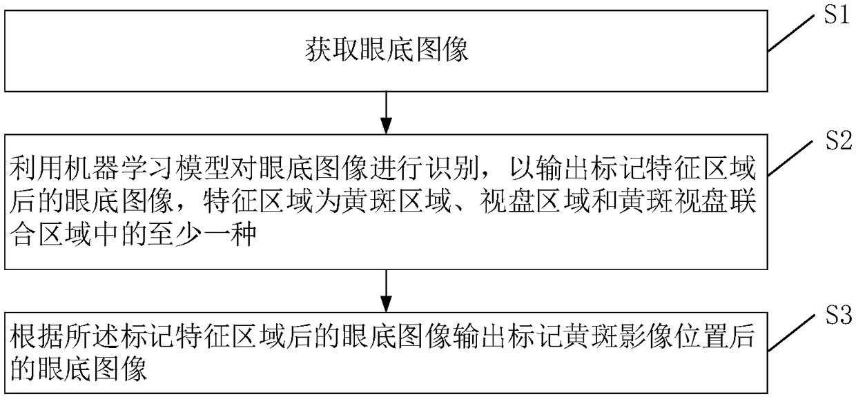
[0042] An embodiment of the present invention provides a macular image detection method, which can be executed by electronic devices such as personal computers and servers, such as figure 2 The method shown includes the following steps:

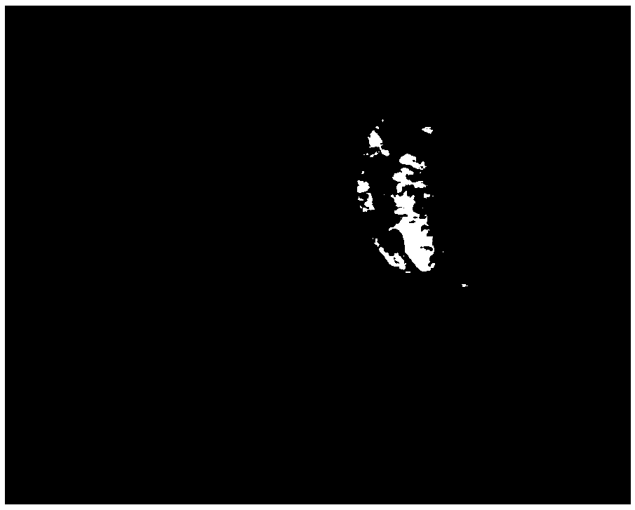
[0043] S1, acquiring fundus images. Such as figure 1 As shown in , ...


PUM


Abstract

The invention provides a macula lutea image detection method and equipment. The macula lutea image detection method comprises the steps that a fundus image is acquired; a machine learning model is utilized to recognize the fundus image to output the fundus image obtained after feature regions are marked, wherein the feature regions are at least one of a macula lutea region, an optic disc region and a macula lutea and optic disc united region, and the machine learning model is obtained by performing training through sample images at the positions of the known feature regions; and according to the fundus image obtained after the feature regions are marked, the fundus image obtained after a macula lutea image position is marked is output.

Description

technical field [0001] The invention relates to the field of medical image processing, in particular to a macular image detection method and equipment. Background technique [0002] In the medical field, the macula is located at 0.35cm on the temporal side of the optic disc of the fundus and slightly below it, in the optical center of the human eye, and is the projection point of the vision axis. The macula is located in the center of the retina, where a large number of visual function cells are concentrated. Abnormalities in the macular area often directly lead to the decline of visual ability. If the lesions in the macular area are not detected and treated in time, the chance of blindness will be greatly increased. [0003] In order to improve the efficiency of doctors observing fundus images, the existing technology proposes to automatically identify and mark the macular area in the fundus image according to the shape, color and other characteristics of the macula throug...

Claims


Application Information

Patent Timeline
Patent Type & Authority Applications(China)
IPC IPC(8): G06T7/00G06N99/00G06K9/00
CPCG06T7/0012G06T2207/20081G06T2207/30041G06T2207/10004G06V40/18
Inventor 王欣熊健皓赵昕罗元元
Owner SHANGHAI EAGLEVISION MEDICAL TECH CO LTD
Why Eureka
Try Eureka