
An image diagnostic device for radiology

A radiology and imaging technology, applied in the field of medical devices, can solve the problems of inconvenience, magnification, and low lamp life.

Inactive Publication Date: 2021-05-11
AFFILIATED YONGCHUAN HOSPITAL OF CHONGQING MEDICAL UNIV
View PDF10 Cites 0 Cited by
  • Summary
  • Abstract
  • Description
  • Claims
  • Application Information

AI Technical Summary

Problems solved by technology

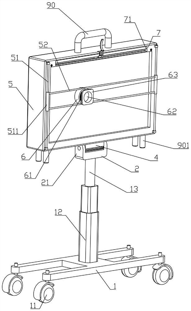
[0004] The purpose of the present invention is to provide a radiological image diagnostic image reading device, which is used to solve the problems of inconvenient use, easy drop of X-ray film, low lamp life and failure to correct the existing diagnostic image reading device. The problem of partial zoom-in viewing

Method used


Image

  • An image diagnostic device for radiology
  • An image diagnostic device for radiology
  • An image diagnostic device for radiology

Examples


Embodiment Construction

[0033] In order to enable those skilled in the art to better understand the present invention, the technical solution of the present invention will be further described below in conjunction with the accompanying drawings and embodiments.

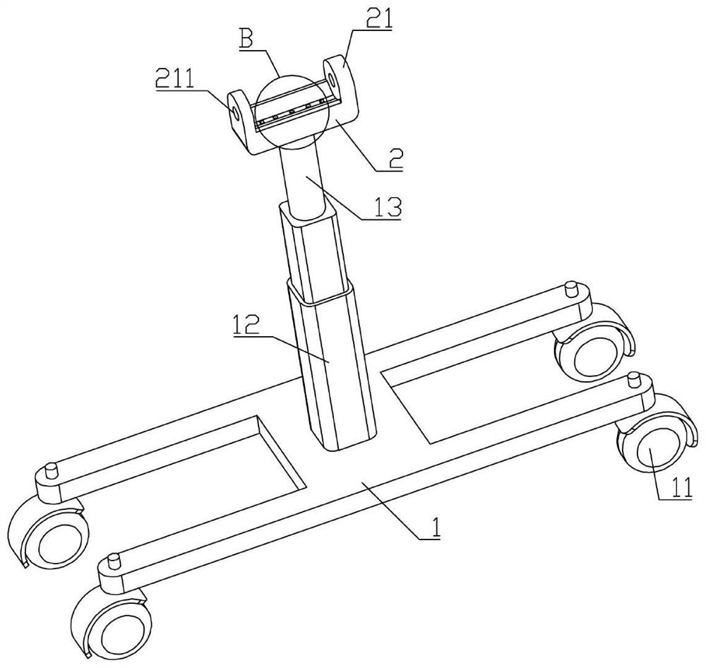
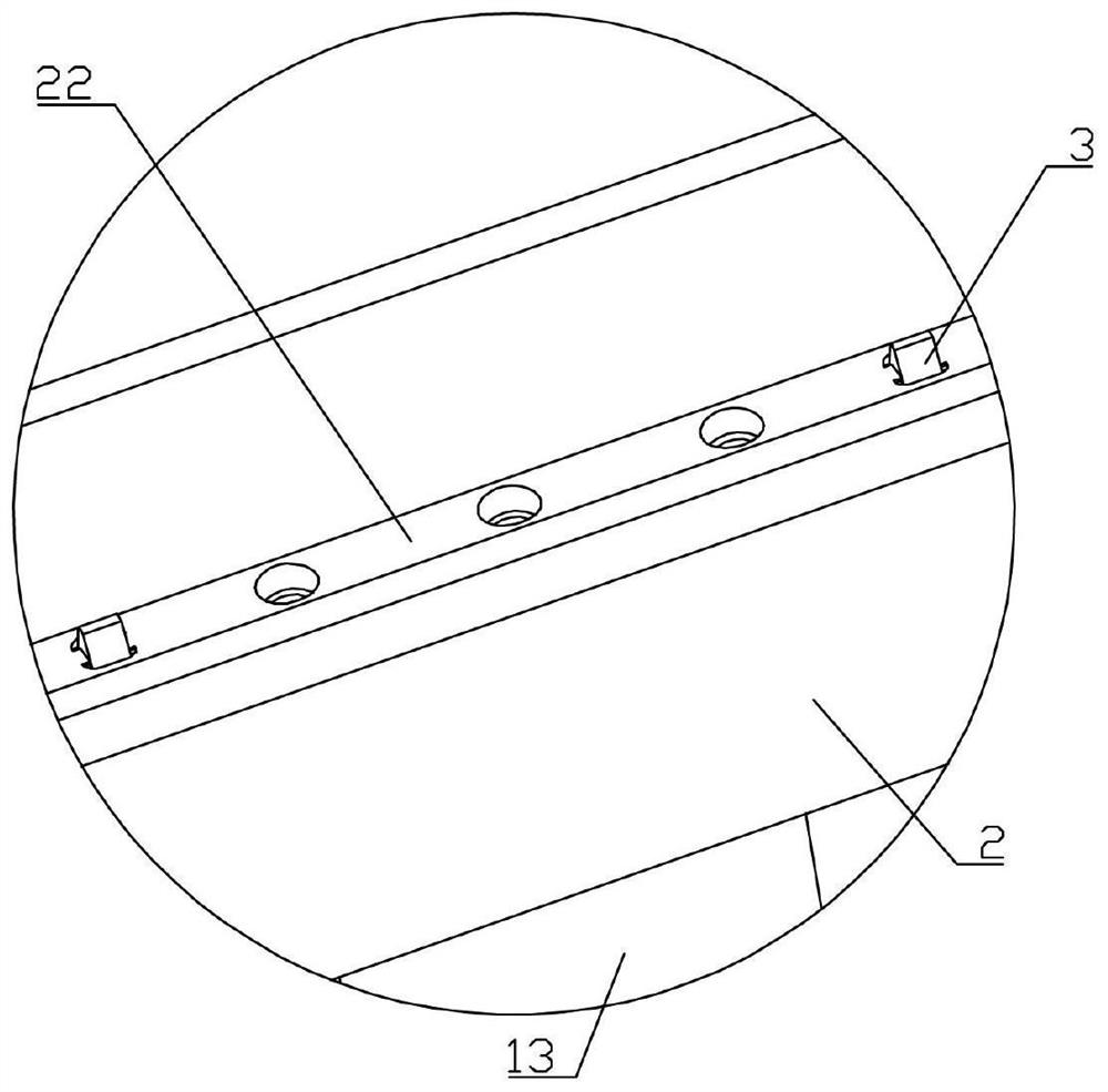
[0034] Such as Figure 1 to Figure 13 As shown, a radiology diagnostic image reading device of the present invention includes an I-shaped base 1, and the lower end surface of the I-shaped base 1 is provided with four universal wheels 11 with a parking lock structure, and the I-shaped base 1 is equipped with lifting Structure 12, the upper end surface of the lifting structure 12 is provided with a first blind hole, a rotating shaft 13 is installed in the first blind hole, a bearing is installed between the rotating shaft 13 and the lifting structure 12, and a connecting block 2 is arranged horizontally on the upper end surface of the rotating shaft 13 , the left and right ends of the upper end surface of the connecting block 2 are integrally ...


PUM


Abstract

The invention belongs to the technical field of medical devices, and in particular relates to a radiological image diagnostic reading device, which comprises an I-shaped base, a sleeve is set on the first sliding rod, and two second sliding rods are arranged between the two sleeves , the outer wall of the bottom cover is provided with a chute matching the second slide bar, the bottom cover is clamped between the two second slide bars through the chute, the inner cover of the bottom cover is provided with a middle cover, and a convex lens is installed in the middle cover, The front end of the strip plate is provided with a through groove, and the transparent plate is provided with a first strip groove at the corresponding position of the through groove. The length of the first strip groove is greater than the length of the through groove. The second bar-shaped slot matched with the bar-shaped slot, a pressing bar is arranged between the first bar-shaped slot and the second bar-shaped slot, and the button switch is installed on the wire between the storage battery and the lamp tube. Its purpose is to solve the problems of inconvenient use, easy drop of X-ray film, low life of lamp tube and inability to zoom in and view parts of the existing diagnostic film reading device caused by structural defects.

Description

technical field [0001] The invention belongs to the technical field of medical devices, and in particular relates to an image diagnosis device for radiology. Background technique [0002] In the imaging diagnosis of the radiology department, patients are often diagnosed with the help of X-ray films made by X-ray equipment, so film reading plays an important role in the diagnosis of diseases and subsequent targeted treatment. The existing film reading device It uses a light box hanging on the wall as a light source, and then puts the image light film on the surface of the light box for reading, but the existing device cannot be moved, and medical personnel need to go to a designated place to read the film, which is very inconvenient, and the height of the light box , The angle cannot be adjusted, and it is inconvenient to use. At the same time, most of the existing X-ray films are directly stuck on the light box when viewed, which is very easy to drop. Moreover, when the ex...

Claims


Application Information

Patent Timeline
Patent Type & Authority Patents(China)
IPC IPC(8): G02B27/02
CPCG02B27/024G02B27/028
Inventor 李德霞
Owner AFFILIATED YONGCHUAN HOSPITAL OF CHONGQING MEDICAL UNIV
Learn More