Method and system for determining sagittal direction medial surface of craniomaxillofacial soft tissue based on optical imaging

A technology for optical imaging and determining methods, applied in image enhancement, image analysis, image data processing, etc.

Active Publication Date: 2018-11-20
AFFILIATED STOMATOLOGICAL HOSPITAL OF NANJING MEDICAL UNIV
View PDF3 Cites 12 Cited by
  • Summary
  • Abstract
  • Description
  • Claims
  • Application Information

AI Technical Summary

Problems solved by technology

[0003] The purpose of the present invention is to provide a method and system for determining the sagittal axis of cranio-maxillofacial soft tissue based on optical imaging, which solves the lack of symmetry in the analysis of facial soft tissue in the existing cranio-maxillofacial analysis methods in the prior art. The problem of determining the axial surface

Method used


Image

  • Method and system for determining sagittal direction medial surface of craniomaxillofacial soft tissue based on optical imaging
  • Method and system for determining sagittal direction medial surface of craniomaxillofacial soft tissue based on optical imaging
  • Method and system for determining sagittal direction medial surface of craniomaxillofacial soft tissue based on optical imaging

Examples


Embodiment

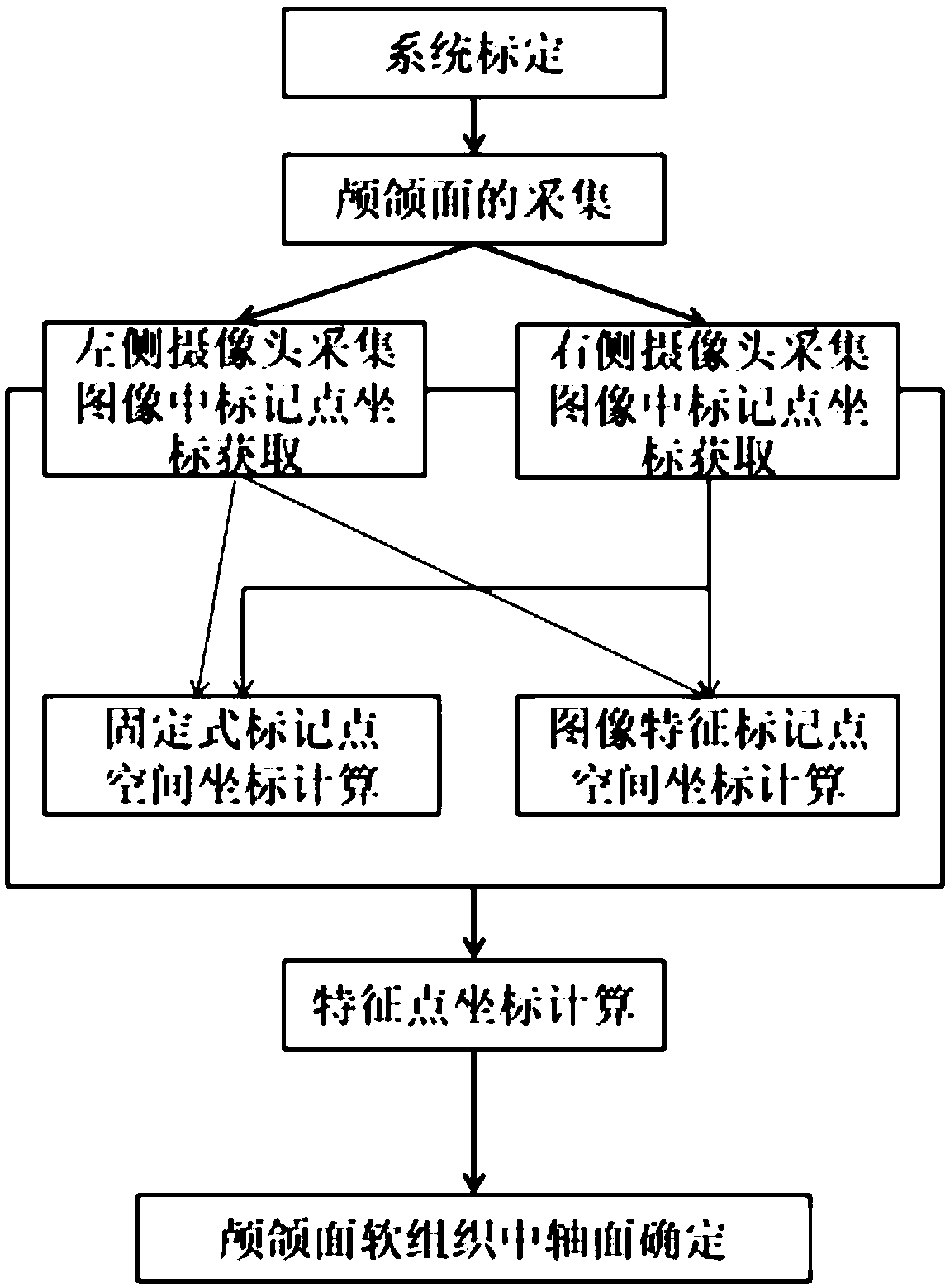
[0042] A method for determining the sagittal axis plane of cranio-maxillofacial soft tissue based on optical imaging, such as figure 1 , including the following steps,

[0043] S1. Three-dimensional space calibration: use Zhang Zhengyou’s camera calibration method in binocular vision technology to calibrate, and obtain the internal and external parameters of the camera group; establish a three-dimensional coordinate system with the midpoint of the two cameras as the origin of the world coordinates: take the camera lens L center point O l with camera lens R center point O r The middle point O of the connection line is the origin of the coordinate system, the line connecting the center of the camera lens is the X axis, which is parallel to the ground, the Y axis is perpendicular to the ground, and the Z axis is perpendicular to the X axis and the Y axis. The acquisition diagram is as follows figure 2 shown;

[0044] S2. Craniomaxillofacial collection: let the subject stand fac...


PUM


Abstract

The invention provides a method and a system for determining a sagittal direction medial surface of a craniomaxillofacial soft tissue based on optical imaging. The method comprises: 1) calibrating three-dimensional space; 2) acquiring facial images; 3) obtaining coordinates of mark points on the acquired image; 4) calculating each characteristic point by using the three-dimensional space coordinates of each mark point, and determining a sagittal direction medial surface. The method and the system can determine the sagittal direction medial surface of a facial soft tissue by each mark point ina three-dimensional face, and determination of the axial plane is the basis of facial soft tissue symmetry analysis and clinical diagnosis analysis related to the facial soft tissue, especially when hard tissue cannot be obtained at the same time. Compared with the prior art, the method and the system can conveniently, effectively and objectively determine the position of the sagittal direction medial surface of craniomaxillofacial soft tissue in a three-dimensional space, especially in a case that hard tissue cannot be obtained.

Description

technical field [0001] The invention relates to a method and system for determining the sagittal axial plane of craniomaxillofacial soft tissue based on optical imaging, which belongs to the field of medical optoelectronic applications. Background technique [0002] The design of traditional computer-aided cranio-maxillofacial surgery system is generally based on cephalometric film in two-dimensional space, and the result is not accurate enough. Relying on 2D cranial analysis systems can improve patient-physician communication, but cannot yet describe it accurately. The optical-based three-dimensional facial imaging technology has been developed in recent years and has gradually been widely used. However, the existing cranio-maxillofacial analysis methods lack methods for determining the axis of symmetry in facial soft tissue analysis. Contents of the invention [0003] The purpose of the present invention is to provide a method and system for determining the sagittal axi...

Claims


Application Information

Patent Timeline
Patent Type & Authority Applications(China)
IPC IPC(8): G06T7/80G06T7/66G06K9/00
CPCG06T7/66G06T7/80G06T2207/30004G06T2207/10012G06V40/166
Inventor 谢理哲胡轶宁王征李维维潘永初吴大明夏阳王林
Owner AFFILIATED STOMATOLOGICAL HOSPITAL OF NANJING MEDICAL UNIV
Why Eureka
Try Eureka