
An annotation extraction method for medical imaging lesions that can improve doctor efficiency

A medical imaging and extraction method technology, applied in the field of radiology, can solve the problems that affect the efficiency and accuracy of doctors' labeling, third-party tools and software may not fully support it, and it is not easy to identify and find the location of lesions, etc., so as to simplify the participation of doctors link, reduce time, and efficiently mark the effect of lesions

Active Publication Date: 2021-10-08
HARBIN INST OF TECH +1
View PDF22 Cites 0 Cited by
  • Summary
  • Abstract
  • Description
  • Claims
  • Application Information

AI Technical Summary

Problems solved by technology

[0006] (2) In the actual diagnosis process, it is not easy to accurately identify and find the location of the lesion
Doctors need to refer to a large amount of historical diagnostic information in the process of labeling, which MITKworkbench cannot provide
This will greatly affect the annotation efficiency and accuracy of doctors
[0007] (3) Third-party tool software may not fully support the data format of a specific hospital
Only CT images are applicable in the annotation process of MITK workbench

Method used


Image

  • An annotation extraction method for medical imaging lesions that can improve doctor efficiency
  • An annotation extraction method for medical imaging lesions that can improve doctor efficiency
  • An annotation extraction method for medical imaging lesions that can improve doctor efficiency

Examples


specific Embodiment approach 2

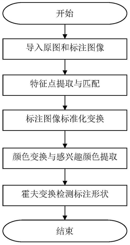
[0058] Specific implementation mode 2: In this implementation mode, the annotation and extraction of metastatic lesions of lung cancer is taken as an example. The doctor uses a drawing tool to draw a red circle in the imaging diagnosis system of the hospital, marks the shape of the lesions and saves the screenshot as a jpg image. The process and main points of implementing the present invention will be described in detail below. The overall execution process follows figure 1 shown.

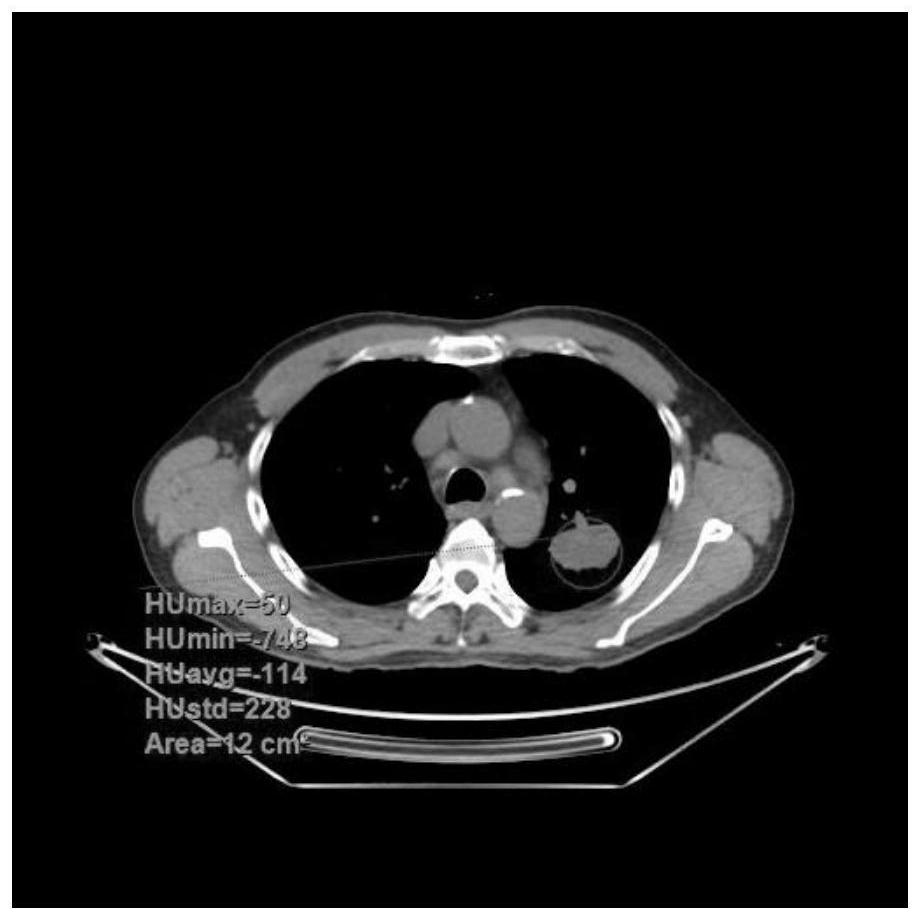
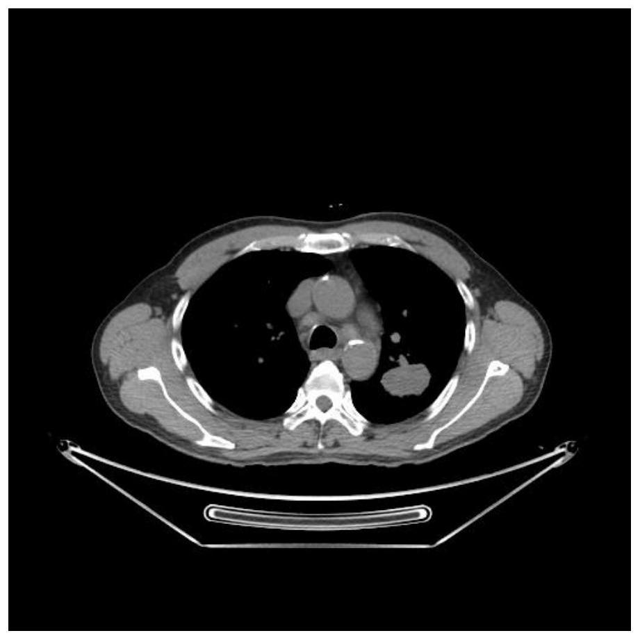
[0059] The first step is to load the original image and the labeled image. Annotated images and original images are shown in figure 2 and 3 shown. When adding a window, the values ​​of TH1 and TH2 in formula (1) are 160 and 240, respectively.

[0060] The second step is feature point extraction and matching. Here the feature points use the SIFT descriptor. The algorithm mainly includes 5 steps for matching:

[0061] 1) Construct scale space, detect extreme points, and obtain scale invaria...


PUM


Abstract

The invention discloses a method for labeling and extracting medical imaging lesions that can improve the efficiency of doctors. The method includes the following steps: 1. Importing the original image dicom image and labeling image; 2. Extracting and matching feature points; 3. Matching the labeling image Carry out standardized transformation; 4. Color transformation and extraction of interesting colors; 5. Label shape detection. The invention allows radiologists to identify and locate lesions on the existing image diagnosis system they are familiar with, and then mark the peripheral shape of lesions, and the subsequent labeling and extraction work is all automated, and image processing technology, feature extraction technology and matching technology are comprehensively used The annotation information of the lesion is restored.

Description

technical field [0001] The invention belongs to the technical field of radiation medicine, and relates to a method for labeling medical image data, in particular to a method for labeling and extracting medical image lesions which can improve the efficiency of doctors. Background technique [0002] In radiology, radiologists usually use CAD (Computer Aided Detection System) such as CT (Computed Tomography) and PET (Positron Emission Computed Tomography) to obtain patient image information. The image information is saved in the dicom format file. In addition to the main pixel information, the dicom file also contains a series of information such as the patient's name, gender, age, image type, and image serial number. The radiologist summarizes the medical imaging information to obtain the findings, draws the patient's diagnosis opinion based on his own experience, and generates a diagnosis report. At present, there has been a lot of research on the automatic recognition of me...

Claims


Application Information

Patent Timeline
Patent Type & Authority Patents(China)
IPC IPC(8): G06T7/00G06T7/73G06K9/46G06K9/62G16H30/40
CPCG06T7/0012G06T7/73G16H30/40G06V10/462G06V10/56G06V10/757G06V2201/03
Inventor 苏统华李彬霍栋
Owner HARBIN INST OF TECH
Learn More