A cervical cell pathological slice classifying method with high and low resolution combination

A high- and low-resolution, pathological section technology, applied in the field of automatic interpretation of cervical cell pathological sections, to achieve the effect of taking into account the accuracy and efficiency, and saving the cost of labeling

Active Publication Date: 2018-12-18
怀光智能科技(武汉)有限公司
View PDF9 Cites 32 Cited by
  • Summary
  • Abstract
  • Description
  • Claims
  • Application Information

AI Technical Summary

Problems solved by technology

Artificially designed pathological cell feature e

Method used


Image

  • A cervical cell pathological slice classifying method with high and low resolution combination
  • A cervical cell pathological slice classifying method with high and low resolution combination
  • A cervical cell pathological slice classifying method with high and low resolution combination

Examples


Embodiment Construction

[0058] In order to make the object, technical solution and advantages of the present invention clearer, the present invention will be further described in detail below in conjunction with the accompanying drawings and embodiments. It should be understood that the specific embodiments described here are only used to explain the present invention, not to limit the present invention. In addition, the technical features involved in the various embodiments of the present invention described below can be combined with each other as long as they do not constitute a conflict with each other.

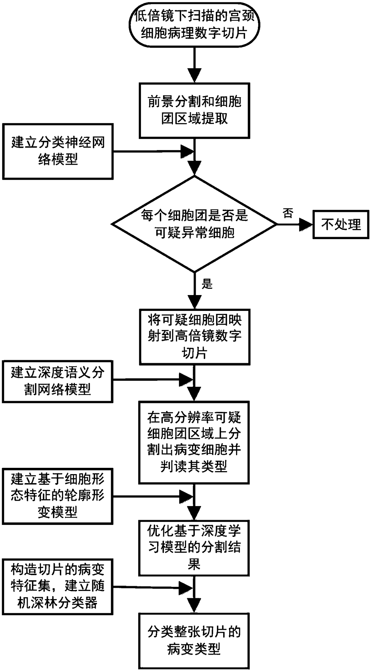
[0059] Such as figure 1 As shown, the method for automatic interpretation of cervical cell pathological sections provided by the present invention comprises the following steps:

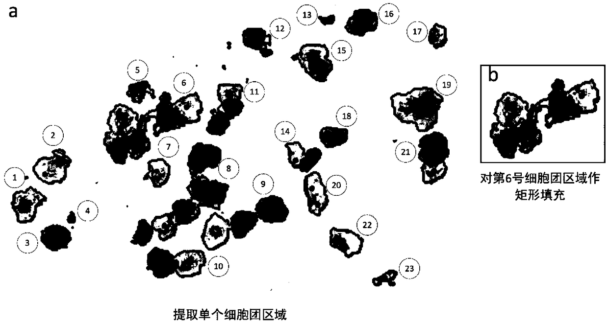
[0060] Step 1) Perform foreground segmentation on the digital slices scanned under a 10x microscope, and extract multiple cell cluster regions in the segmented foreground image. The collection of cells connected by c...


PUM


Abstract

The invention discloses a cervical cell pathological slice automatic judging method with high and low resolution combination, which is characterized in that the method comprises the following steps: extracting a single cell mass region on a low-resolution cervical pathological slice image; extracting a single cell mass region from the low-resolution cervical pathological slice image; identifying suspicious abnormal cell clusters from low-resolution cell clusters; mapping suspected abnormal cell mass regions into high resolution slice images; semantically segmenting pathological cells from thehigh-resolution region of suspicious cell clusters and interpreting the type; according to the type, quantity and confidence of the segmented lesion cells, establishing the feature set of the slice, and then classifying the lesion type of the whole slice. The invention takes cell cluster as processing and recognition unit, utilizes classification neural network model to quickly identify suspiciousabnormal cell cluster on low-resolution digital slice, then divides out pathological cell on high-resolution suspicious area and judges its type, at the same time, improves recognition accuracy and recognition efficiency.

Description

technical field [0001] The invention belongs to the field of medical cytopathological image processing, and more specifically relates to an automatic interpretation method for cervical cytopathological slices combined with high and low resolution. Background technique [0002] Cervical cancer is a malignant tumor with high incidence in women. Cervical liquid-based cytopathology is currently the most important means of preventing and screening cervical cancer. Accurate interpretation of diseased cells in cytopathological slice images is an important basis for doctors to determine the patient's condition and formulate a treatment plan. At present, the manual interpretation of cytopathological images is not only time-consuming, but also the interpretation results are very dependent on the experience of doctors. Therefore, automatically interpreting diseased cells in pathological slides can not only improve the efficiency of diagnosis but also provide doctors with a more unifi...

Claims


Application Information

Patent Timeline
IPC IPC(8): G06K9/62G06K9/34G06K9/32
CPCG06V10/25G06V10/267G06V2201/03G06F18/24
Inventor 程胜华余江胜曾绍群刘秀丽吕晓华
Owner 怀光智能科技(武汉)有限公司
Why Eureka
Try Eureka