A method and apparatus for processing cervical cytological image features

A technology of image features and processing methods, which is applied in the field of processing methods and devices for cervical cytology image features, and can solve problems such as poor results and large differences

Pending Publication Date: 2018-12-18
马丁
View PDF4 Cites 17 Cited by
  • Summary
  • Abstract
  • Description
  • Claims
  • Application Information

AI Technical Summary

Problems solved by technology

[0006] However, due to the large difference between the segmentation of medical images and natural images, it is often not effective to...

Method used


Image

  • A method and apparatus for processing cervical cytological image features
  • A method and apparatus for processing cervical cytological image features
  • A method and apparatus for processing cervical cytological image features

Examples


Embodiment 1

[0096] Embodiment 1. A method for feature processing of cervical cytology images, comprising:

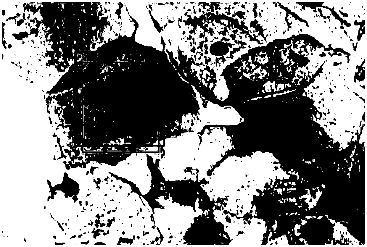
[0097] (1) Prepare a 40-fold magnified cervical cytology image and an annotation frame of unconventional cells in the image as training data; each annotation frame includes the abscissa and ordinate of the upper left corner of the frame, the width and height of the frame, and the The category corresponding to the box; the classification category corresponding to the box includes high-grade squamous epithelial lesion, low-grade squamous epithelial lesion, atypical squamous cell and squamous cell carcinoma, etc.;

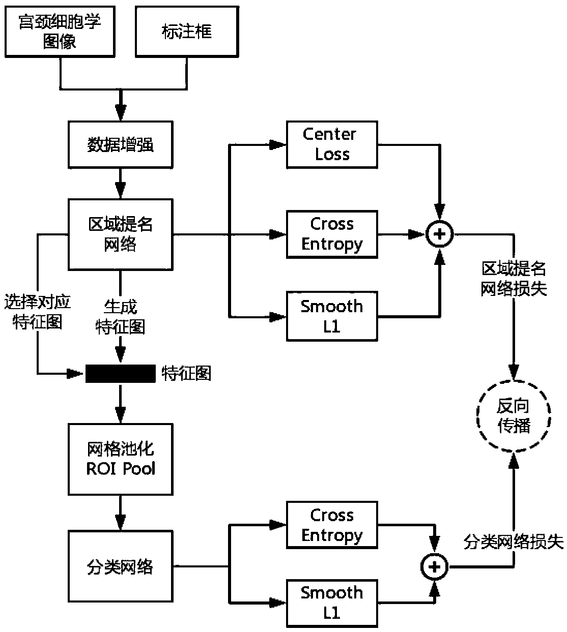
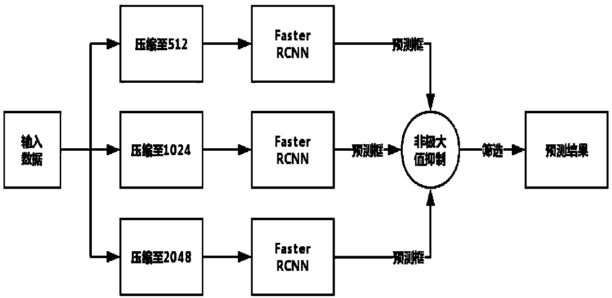
[0098] (2) Compress the training data obtained in step (1) to the resolution R, and input the region nomination network based on ResNet after the cervical cytology image data is enhanced, to obtain the region nomination frame and cervical cytology image feature map;

[0099] The specific steps of the data enhancement method are as follows:

[0100] (2-1) Flip the image and...

Embodiment 2

[0133] Embodiment 2, a method for feature processing of cervical cytology images, comprising:

[0134] (1) Prepare a 20-fold magnified cervical cytology image and an annotation frame of unconventional cells in the image as training data; each annotation frame includes the abscissa and ordinate of the upper left corner of the frame, the width and height of the frame, and the The category corresponding to the box; the classification category corresponding to the box includes high-grade squamous epithelial lesion, low-grade squamous epithelial lesion, atypical squamous cell and squamous cell carcinoma;

[0135] (2) Compress the training data obtained in step (1) to the resolution R, and input the region nomination network based on ResNet after the cervical cytology image data is enhanced, to obtain the region nomination frame and cervical cytology image feature map;

[0136] The specific steps of the data enhancement method are as follows:

[0137] (2-1) Flip the image and label...


PUM


Abstract

The invention discloses a method and apparatus for processing cervical cytological image features. The cervical cytology image data is compressed to different resolutions and input into a region nomination network to obtain a region nomination frame and a cervical cytology image characteristic map. In the cervical cytology image feature map, the feature corresponding to the region nomination box is selected as input, and the pool feature map is obtained through the grid pool layer. The classification probability of the region and the deviation between the prediction box and the nomination boxare obtained by inputting the pooled characteristic map into the classification network. The loss function is obtained by calculating the loss of the regional nomination network and the loss of the classification network. The convergent Faster RCNN model is obtained by back propagation optimization. Finally, a non-maximum suppression method is used to screen Faster RCNN trained by images with different resolutions to obtain a final prediction frame. The invention can effectively improve the efficiency and accuracy of screening unconventional cells in cervical cytology images by doctors.

Description

technical field [0001] The invention belongs to the field of medical image data processing, in particular to a method and device for processing cervical cytology image features. Background technique [0002] In the clinical diagnosis of cervical cancer, the results of pathological diagnosis are considered to be the most authoritative and accurate discrimination results, and are also the most important indicators for clinical diagnosis of cancer. In the pathological cytology imaging of cervical cancer, clinicians can scan the entire slice with the naked eye by a professional pathologist through the movement of the slice under the microscope, and find no intraepithelial lesion / malignant lesion cells (NILM), low Unconventional cells such as high-grade squamous intraepithelial lesion (LSIL) and high-grade squamous intraepithelial lesion (HSIL) are tedious and time-consuming for experienced doctors, and with the increase of reading time , the misdiagnosis rate also increased. ...

Claims


Application Information

Patent Timeline
IPC IPC(8): G06K9/62
CPCG06V2201/03G06F18/2415G06F18/214
Inventor 马丁吴健黄晓园王彦杰
Owner 马丁
Why Eureka
Try Eureka