A superpixel method for medical image segmentation

A medical image and super pixel technology, applied in the field of medical image, can solve the problem of the influence of positioning accuracy

Active Publication Date: 2018-12-18
SHANDONG UNIV OF FINANCE & ECONOMICS
View PDF7 Cites 62 Cited by
  • Summary
  • Abstract
  • Description
  • Claims
  • Application Information

AI Technical Summary

Problems solved by technology

Although the effect is good, the trade-off between the layer and the size of the receptive field has a great impact on the positioning accuracy

Method used


Image

  • A superpixel method for medical image segmentation
  • A superpixel method for medical image segmentation
  • A superpixel method for medical image segmentation

Examples


Embodiment Construction

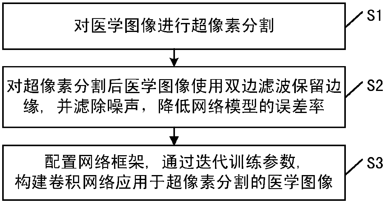
[0070] The present invention provides a superpixel method for medical image segmentation, such as figure 1 shown, methods include:

[0071] S1, perform superpixel segmentation on medical images;

[0072] S2, use bilateral filtering to preserve the edges of the medical image after superpixel segmentation, and filter out noise to reduce the error rate of the network model;

[0073] S3, configure the network framework, and construct a convolutional network for superpixel segmentation of medical images by iteratively training parameters.

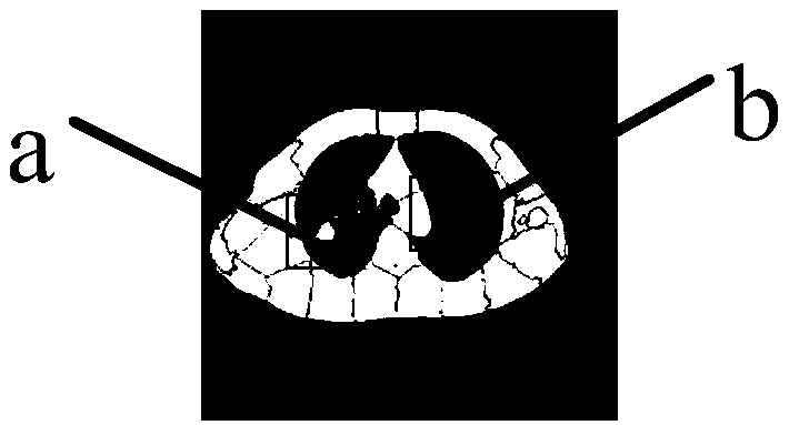
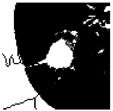
[0074] Image segmentation is an important branch of analyzing and identifying the semantic information of medical images, and superpixel-level image processing is a simple and effective method. However, due to the intricate distribution of tissues in medical images, the superpixel segmentation results are blurred in the edge information part, and the cascading updates of each category of segmentation results are obvious. To this end, the pres...


PUM


Abstract

The invention provides a superpixel method for medical image segmentation. The method comprises the steps of: processing a medical image into a superpixel; for the medical images obtained after superpixel segmentation, using bilateral filtering to preserve the edge and filter the noise to reduce the error rate of the network model; configuring a network framework, and constructing a convolution network for the medical images obtained after superpixel segmentation by iterative training parameters. Based on the linear iterative clustering segmentation method, this method applies the thought of the U-Net network to the post-optimization of super-pixels, which makes up the defect of inaccurate segmentation of inner edge of super-pixel, increases the standard layer to improve the weight sensitivity of each network layer, improves the convergence performance of the network, and makes the segmentation result closer to the actual value. Because the anatomical structure and pathological tissueof medical images are very clear, the medical images segmented by SLIC algorithm can obtain more comprehensive super-pixel, and the edge accuracy of super-pixel can be further improved by convolutionnetwork.

Description

technical field [0001] The invention relates to the field of medical images, in particular to a superpixel method for medical image segmentation. Background technique [0002] Medical images from various imaging techniques [1] , such as ultrasound, computed tomography (CT), X-ray, and magnetic resonance imaging (MRI), are used to describe the anatomical structure of different tissues of the human body. The anatomical map depicted by medical images reflects the health of the human body, and understanding the detailed division of regions in the anatomical map of various parts of the human body is helpful for auxiliary diagnosis and next-step treatment. For example, the lung window includes the lung parenchyma, mediastinum, pleura, and diseased tumors. Accurate segmentation of tumors can help determine the condition more comprehensively and accurately, select appropriate radiotherapy methods, and improve the success rate of treatment. However, the traditional manual segmentat...

Claims


Application Information

Patent Timeline
Patent Type & Authority Applications(China)
IPC IPC(8): G06T7/10
Inventor 王海鸥刘慧郭强张小峰高珊珊姜迪
Owner SHANDONG UNIV OF FINANCE & ECONOMICS
Why Eureka
Try Eureka