
X-ray auxiliary photographing method based on infrared camera, system and device

An infrared camera and photography device technology, applied in the field of medical radiation, can solve the problems of inaccurate tilt angle, high retake rate, and body stiffness, etc., and achieve the effects of improving work efficiency, reducing radiation dose, and reducing retake rate

Active Publication Date: 2019-01-04
NANFANG HOSPITAL OF SOUTHERN MEDICAL UNIV
View PDF8 Cites 4 Cited by
  • Summary
  • Abstract
  • Description
  • Claims
  • Application Information

AI Technical Summary

Problems solved by technology

In actual operation, due to individual differences, technicians do not have a quantitative standard for the tilt angle of the patient's pelvis when taking pictures, and the tilt angle required by male and female subjects is also different, so it is easy to cause inaccurate tilt angle, position Incorrect, so that the photos taken cannot meet the diagnostic requirements, and often need to be re-taken, the efficiency is low, and unnecessary radiation doses are added to the patient
[0004] In addition, some subjects, especially those with ankylosing spondylitis, are relatively stiff when shooting the double oblique view of the sacroiliac joint, and often cannot persist in cooperating to complete the entire shooting
During the shooting process, abnormal situations such as body movement are prone to occur, which will also lead to blurred images, poor display of articular surfaces, failure to achieve diagnostic purposes, and high retake rate
And if the family members are asked to help fix the subject, even though they wear protective clothing, the family members will still passively receive additional radiation doses

Method used


Image

  • X-ray auxiliary photographing method based on infrared camera, system and device
  • X-ray auxiliary photographing method based on infrared camera, system and device
  • X-ray auxiliary photographing method based on infrared camera, system and device

Examples


Embodiment Construction

[0045] The present invention will be further explained and described below in conjunction with the accompanying drawings and specific embodiments of the description. For the step numbers in the embodiment of the present invention, it is only set for the convenience of explanation and description, and there is no limitation on the order of the steps. The execution order of each step in the embodiment can be carried out according to the understanding of those skilled in the art Adaptive adjustment.

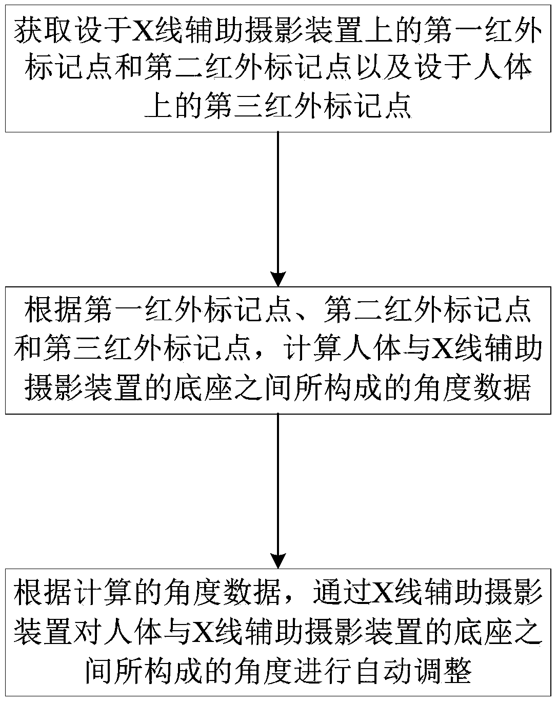
[0046] refer to figure 1 , the present invention is a kind of X-ray auxiliary photography method based on infrared camera, comprises the following steps:

[0047] Obtaining the first infrared marker point and the second infrared marker point set on the X-ray assisted photography device and the third infrared marker point set on the human body;

[0048] According to the first infrared marker point, the second infrared marker point and the third infrared marker point, calculate the ...


PUM


Abstract

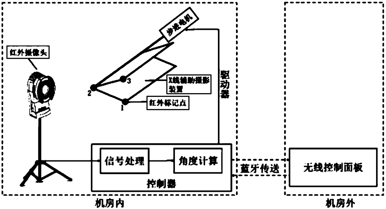
The invention discloses an X-ray auxiliary photographing method based on an infrared camera, a system and a device. The method comprises the following steps of obtaining a first infrared mark point and a second infrared mark point arranged on an X-ray auxiliary photographing device and a third infrared mark point arranged on a human body; calculating angle data constituted between the human body and the base of the X-ray auxiliary photographing device according to the first infrared mark point, the second infrared mark point and the third infrared mark point; according to the calculated angledata, automatically adjusting the angle formed between the human body and the base of the X-ray auxiliary photographing device by the X-ray auxiliary photographing device. The system includes a coordinate obtaining module, an angle calculating module and an angle adjusting module. The apparatus includes a memory and a processor. The invention does not require manual adjustment by technical personnel, improves work efficiency and reduces reshooting rate, further reduces radiation doses received by patients and family members, and can be widely applied in the medical radiation technical field.

Description

technical field [0001] The invention relates to the technical field of medical radiation, in particular to an infrared camera-based X-ray assisted photography method, system and device. Background technique [0002] Clinically, there are more than 20 diseases related to sacroiliac joints, among which ankylosing spondylitis (AS) and osteitis iliacs are the most common, and the diagnosis of sacroiliac joint diseases is generally based on the Imaging data are used as one of the basis for diagnosis. [0003] In the radiology department, the oblique position of the sacroiliac joint is taken. The subject needs to lie supine on the imaging table, hold the head with both hands, raise the subject side to an angle of 20-25°, bend the lower limbs slightly, and the lower edge of the flat panel detector is flat on the pubic bone. edge. Obtain X-ray images and observe the tangent position of the sacroiliac joint on one side to determine whether there are lesions in the bone density, art...

Claims


Application Information

Patent Timeline
IPC IPC(8): A61B6/04A61B6/00
CPCA61B6/54A61B6/0487
Inventor 马乐林晓佳秦耿耿蔡裕兴陈卫国
Owner NANFANG HOSPITAL OF SOUTHERN MEDICAL UNIV
Learn More