Method and device for removing CT (computed tomography) image noises

A CT image and image technology, applied in the field of image denoising, can solve problems such as loss of image edges and contrast, and inability to effectively remove high-frequency noise in CT images.

Active Publication Date: 2013-07-03
GE MEDICAL SYST GLOBAL TECH CO LLC
View PDF2 Cites 30 Cited by
  • Summary
  • Abstract
  • Description
  • Claims
  • Application Information

AI Technical Summary

Problems solved by technology

[0004] Aiming at the problem that the prior art cannot effectively remove the high-frequency noise of the CT image, and will lose the image edg

Method used


Image

  • Method and device for removing CT (computed tomography) image noises
  • Method and device for removing CT (computed tomography) image noises
  • Method and device for removing CT (computed tomography) image noises

Examples


Embodiment Construction

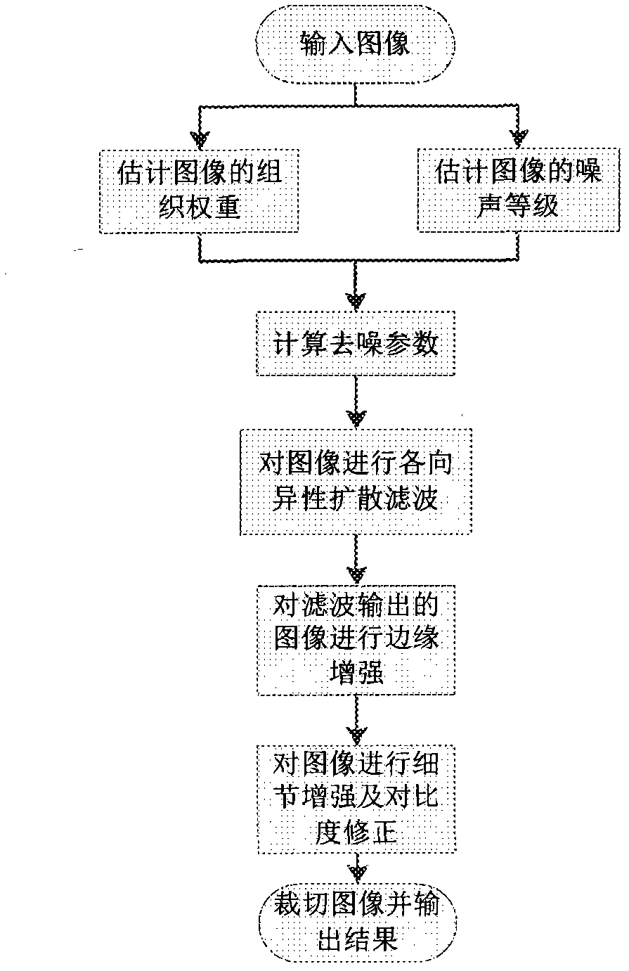
[0101] The present invention will be further described below in conjunction with the accompanying drawings and specific embodiments.

[0102] This embodiment discloses a method for removing CT image noise, including the following steps:

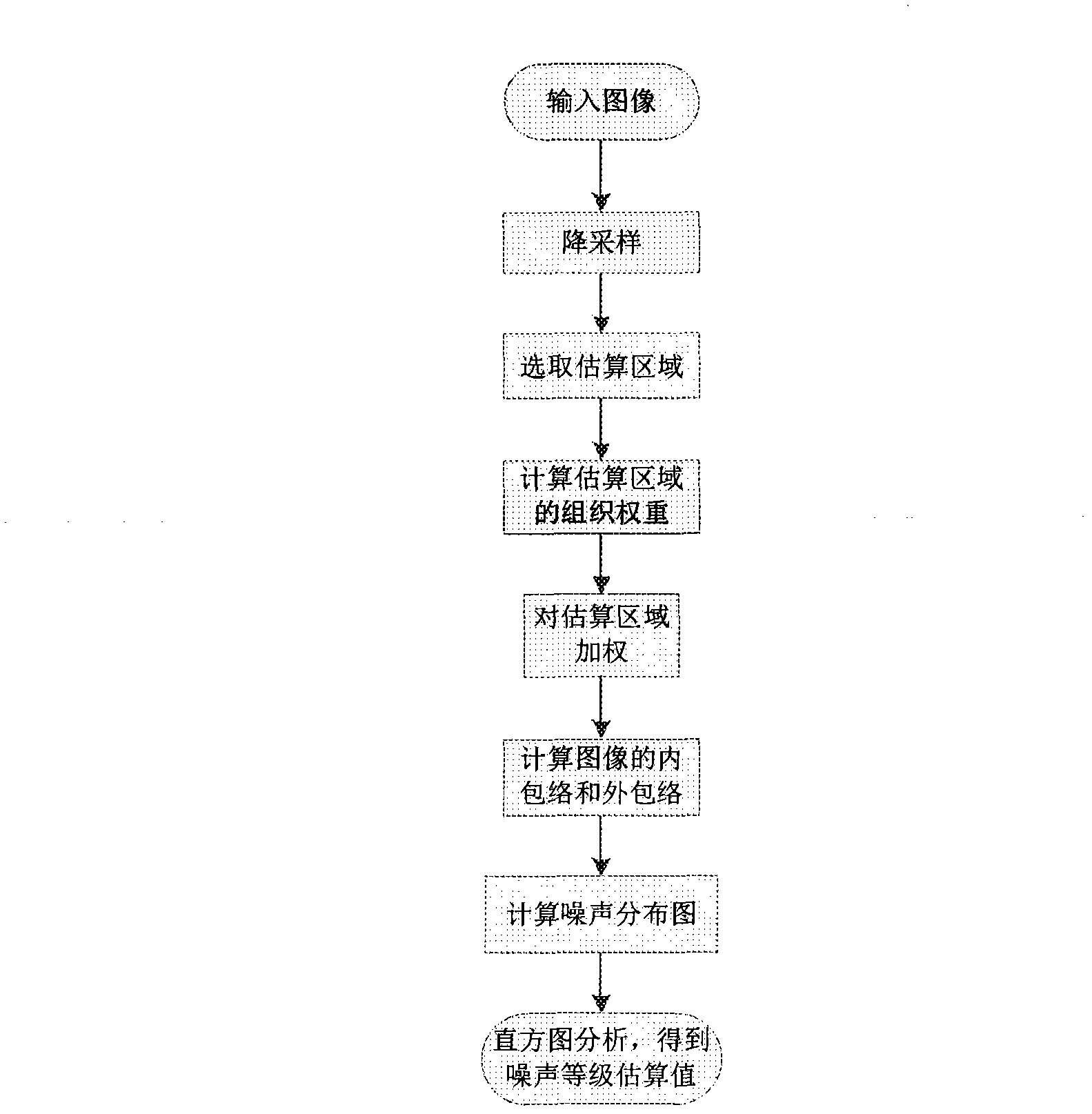
[0103]1. Estimate tissue weights for images

[0104] First configure the initial parameters: tissue mean Tissue, soft tissue first threshold TissueUp, soft tissue second threshold TissueDown, tissue noise reduction weight SRange, average filter window width K;

[0105] Then, within the scope of the display field of view (DFOV), smooth the input image with a smoothing width of K to obtain the smoothing value of each pixel:

[0106]

[0107] Among them, InputImage(i, j) represents the pixel value of the original input image;

[0108] Finally, calculate the tissue weight of each pixel of the input image:

[0109] TissueWeight ( i , j ) = ...


PUM


Abstract

The invention discloses a method and a device for removing CT (computed tomography) image noises, and relates to the technical field of image noise removal. The method comprises the following steps of: estimating the tissue weight of an image, estimating the noise level of the image, calculating a noise removing parameter, carrying out anisotropic diffusion filtering on the image, carrying out edge enhancement on the image subjected to filtering output, enhancing details of the image and correcting the contrast ratio, cutting the image, and outputting the result. The device comprises a module for estimating the tissue weight of the image, a module for estimating the noise level of the image, a module for calculating the noise removing parameter, a module for carrying out anisotropic diffusion filtering on the image, a module for carrying out edge enhancement on the image subjected to filtering output, a module for performing detail enhancement and contrast ratio rectification on the image, and a module for cutting images and outputting the result. The method and the device can maintain the image edge and the original contrast ratio of the image while effectively removing the high-frequency noises of the CT images.

Description

technical field [0001] The invention relates to the technical field of image denoising, in particular to a method and device for removing CT image noise. Background technique [0002] Computed tomography (CT) is a mature and clinically recognized examination method, and its radiation dose has attracted more and more attention. In the current CT, the CT dose is often reduced by reducing the tube current and tube voltage, but this will increase the blocky and granular noise of the image, reduce the image quality, and affect the diagnosis of clinicians. [0003] The current methods for improving the image quality of low-dose CT are mainly divided into two categories: projection space data-based and image space data-based methods. The multi-scale analysis of anisotropic filter and wavelet is a nonlinear adaptive filtering noise reduction method based on image space data. Anisotropic filter can effectively suppress the low-frequency and small gradient noise in CT images. However...

Claims


Application Information

Patent Timeline
IPC IPC(8): G06T5/00
Inventor 董淑琴H·江童晓蕾王斌
Owner GE MEDICAL SYST GLOBAL TECH CO LLC
Why Eureka
Try Eureka