Supercharge Your Innovation With Domain-Expert AI Agents!

A medical image fusion method and system

A medical image and fusion method technology, applied in the field of medical image fusion methods and systems, can solve problems such as small computational complexity, loss of information such as stitching traces, edges, and contours, and achieve comprehensive information, promotion effects, and enhancement of details and clarity. Effect

Active Publication Date: 2021-08-13
GUANGDONG UNIV OF TECH
View PDF2 Cites 0 Cited by
  • Summary
  • Abstract
  • Description
  • Claims
  • Application Information

AI Technical Summary

Problems solved by technology

Image fusion based on wavelet transform: perform wavelet transform on the source image, decompose it in different feature domains of different frequency bands, and then perform fusion in different feature domains, and then use wavelet inverse transform to obtain the fusion image process, but due to wavelet The transformation has no displacement invariance, and if the images are not well registered, the fusion image will be distorted
In addition, the weighted average method is also one of the commonly used fusion methods for medical image fusion. The weighted average method is a weighted average of the pixel gray values ​​corresponding to each source image. Its advantage is that the calculation amount is small, but there are inevitably A certain problem, the ordinary weighted average method is easy to lose information such as edges and contours in the image, and in the case of large differences in the grayscale of the fusion source image, it is easy to appear obvious stitching traces

Method used


Image

  • A medical image fusion method and system
  • A medical image fusion method and system
  • A medical image fusion method and system

Examples


Embodiment 1

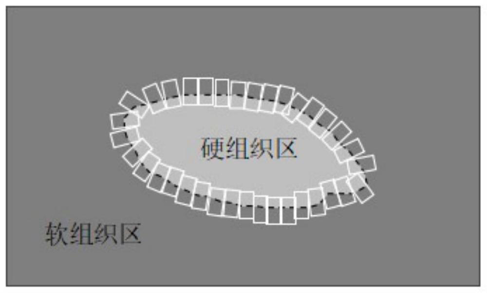
[0046] Based on CT images, the imaging effect of hard tissue is good, but the imaging effect of soft tissue is poor, while ultrasound is the opposite, the imaging effect of soft tissue is good, and the imaging effect of hard tissue is poor. Therefore, the inventor thinks that CT images and ultrasound images can be fully utilized to have different imaging advantages in different local areas, and thus think that a certain area feature of the image can be used as a measuring standard. Therefore, during image fusion, a larger weight should be assigned to the image to be fused with a larger feature advantage, that is, for hard tissue parts, a larger weight is assigned to the CT image, and the weight of the ultrasound image is correspondingly smaller ; and for soft tissue parts, a larger weight is assigned to the ultrasound image, and the weight of the CT image is correspondingly smaller; and appropriate weights are assigned to the CT image and the ultrasound image in the boundary ar...

Embodiment 2

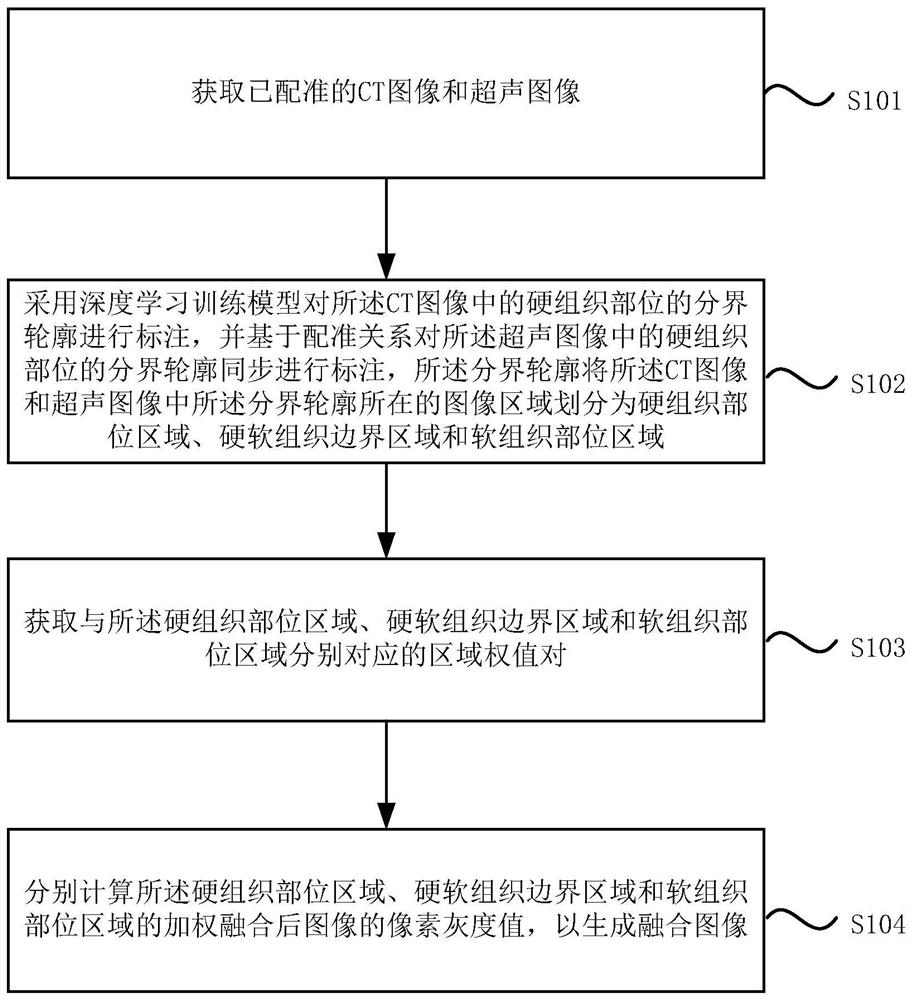
[0066] Please refer to the attached Figure 5 , is a schematic structural diagram of a medical image fusion system provided in Embodiment 2 of the present invention, and the system is suitable for implementing the medical image fusion method provided in the embodiment of the present invention. The system specifically includes the following modules:

[0067] An image acquisition module 21, configured to acquire registered CT images and ultrasound images;

[0068] The contour labeling module 22 is configured to use a deep learning training model to mark the boundary contour of the hard tissue part in the CT image, and to simultaneously mark the boundary contour of the hard tissue part in the ultrasound image based on the registration relationship, The boundary contour divides the image area where the boundary contour is located in the CT image and the ultrasound image into a hard tissue area, a hard and soft tissue boundary area, and a soft tissue area; wherein, the hard and so...


PUM


Abstract

The invention discloses a medical image fusion method and system. The method includes: acquiring a registered CT image and an ultrasonic image; and ultrasonic images are divided into hard tissue area, hard and soft tissue boundary area and soft tissue area; obtain the area weight pairs corresponding to hard tissue area, hard and soft tissue boundary area and soft tissue area respectively ( 1 ,μ 2 ); respectively calculate the pixel gray value X of the weighted fused image of the hard tissue region, hard and soft tissue boundary region, and soft tissue region to generate a fused image. The technical solution provided by the embodiment of the present invention has displacement invariance, and will not distort the fused image when the image registration is not accurate enough, which is conducive to application and promotion, and there will be no obvious splicing traces and loss of edge contour information, which enhances The details and clarity of the image make the information of the fused image more comprehensive and accurate.

Description

technical field [0001] Embodiments of the present invention relate to the technical field of medical image processing, in particular to a medical image fusion method and system. Background technique [0002] Nuclear medicine equipment (mainly including positron emission tomography imaging equipment PET and magnetic resonance imaging equipment MRI) and X-ray computed tomography equipment CT obtain patients' functional and anatomical images for disease diagnosis respectively, and are commonly used in modern imaging diagnostic medicine. two imaging modes. With the rapid development of medical technology and the continuous advancement of nuclear medicine equipment technology, the mainstream nuclear medicine equipment currently on the market is multimodal fusion, mainly PET / CT and MRI / CT. Due to the relatively poor spatial resolution of nuclear medicine equipment, CT images are needed for positioning, etc. Clinicians need to fuse nuclear medicine images and CT images for medical...

Claims


Application Information

Patent Timeline
Patent Type & Authority Patents(China)
IPC IPC(8): G06T11/00G06T5/50
CPCG06T5/50G06T11/008G06T2207/10081G06T2207/10132G06T2207/20221
Inventor 方桂标徐圣兵林森林陈广诚杜钦涛
Owner GUANGDONG UNIV OF TECH
Features
  • R&D
  • Intellectual Property
  • Life Sciences
  • Materials
  • Tech Scout
  • Unparalleled Data Quality
  • Higher Quality Content
  • 60% Fewer Hallucinations
Learn More