Method and device for detecting tumor cells in pleural fluid fluorescence image

A fluorescence image and tumor cell technology, applied in measuring devices, analysis using fluorescence emission, particle and sedimentation analysis, etc., can solve problems such as poor reproducibility, strong subjectivity, large errors, etc., and achieve simple and clear algorithms, Low hardware configuration requirements and good reproducibility

Inactive Publication Date: 2019-04-02
北京羽医甘蓝信息技术有限公司
View PDF6 Cites 2 Cited by
  • Summary
  • Abstract
  • Description
  • Claims
  • Application Information

AI Technical Summary

Problems solved by technology

[0003] In view of this, the present invention provides a method and device for detecting tumor cells in pleural effusion fluorescence images, which can solve the technical problems of low efficiency, strong subjectivity, large errors, and poor reproducibility in the prior art

Method used


Image

  • Method and device for detecting tumor cells in pleural fluid fluorescence image
  • Method and device for detecting tumor cells in pleural fluid fluorescence image
  • Method and device for detecting tumor cells in pleural fluid fluorescence image

Examples


Embodiment Construction

[0026] The following describes exemplary embodiments of the present invention with reference to the accompanying drawings, which include various details of the embodiments of the present invention to facilitate understanding, and should be regarded as merely exemplary. Therefore, those of ordinary skill in the art should realize that various changes and modifications can be made to the embodiments described herein without departing from the scope and spirit of the present invention. Likewise, for clarity and conciseness, descriptions of well-known functions and structures are omitted in the following description.

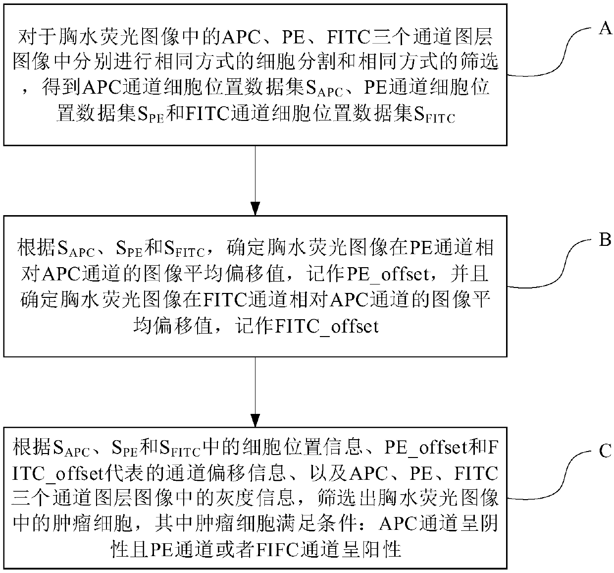
[0027] figure 1 It is a schematic flow chart of a method for examining tumor cells in a fluorescent image of pleural fluid according to an embodiment of the present invention. Such as figure 1 As shown, the method can include the following steps A to C.

[0028] Step A: Perform the same method of cell segmentation and the same method of screening in the three channel ...


PUM


Abstract

The invention provides a method and a device for detecting tumor cells in a pleural fluid fluorescence image. The method comprises the following steps: respectively performing cell segmentation in a same way and screening in a same way on APC, PE and FITC channel layer images in the pleural fluid fluorescence image to obtain a cell position data set SAPC of the APC channel, a cell position data set SPE of the PE channel, and a cell position data set SFITC of the FITC channel; determining an image average offset value of the pleural fluid fluorescence image in the PE channel relative to the APCchannel according to the SAPC, the SPE and the SFITC, marking as PE_offset, and determining the image average offset value of the pleural fluid fluorescence image in the FITC channel relative to theAPC channel, marking as FITC_offset; and screening the tumor cells in the pleural fluid fluorescence image according to the cell position information in the SAPC, the SPE and the SFITC, channel offsetinformation represented by the PE_offset and the FITC_offset, and gray information in the APC, the PE and the FITC channel layer images, wherein the tumor cells satisfy the following conditions: theAPC channel is negative and the PE channel or the FIFC channel is positive.

Description

Technical field [0001] The invention relates to the technical field of computers and software, in particular to a method and a device for detecting tumor cells in fluorescent images of pleural fluid. Background technique [0002] In the field of pathology, it is usually necessary to analyze and detect the fluorescence images of pleural fluid. There are three channels of common pleural fluid fluorescence images, namely APC, PE, and FITC. The maximum absorption of APC is 630nm-650nm, the emission wavelength is 660nm, and the color is roughly red. The maximum absorption of PE is 490nm-560nm, the emission wavelength is 574nm, and the color is roughly orange-yellow. FITC has a maximum absorption wavelength of 488nm and a maximum emission wavelength of 530nm. The color is roughly green. Each channel uses a different fluorescent substance. The total number of cells in a pleural fluid fluorescence image is usually between 20,000 and 200,000, while the number of positive tumor cells (c...

Claims


Application Information

Patent Timeline
Patent Type & Authority Applications(China)
IPC IPC(8): G01N15/00G01N15/10A61B5/00
CPCA61B5/0071G01N15/00G01N15/10G01N15/1056G01N2015/0065G01N2015/1006G01N2015/1081
Inventor 徐子能白海龙丁鹏
Owner 北京羽医甘蓝信息技术有限公司
Why Eureka
Try Eureka