A multi-channel head magnetic resonance imaging tissue segmentation method

A magnetic resonance imaging, multi-channel technology, applied in the field of medical image processing, can solve the problem of not using the anatomical structure information of the brain, and achieve the effect of promoting accurate segmentation, simple method, and improving segmentation accuracy

Active Publication Date: 2019-04-26
SHENZHEN GRADUATE SCHOOL TSINGHUA UNIV
View PDF4 Cites 7 Cited by
  • Summary
  • Abstract
  • Description
  • Claims
  • Application Information

AI Technical Summary

Problems solved by technology

However, the existing deep learning methods do not use the relatively fixed anatomical structure information of

Method used


Image

  • A multi-channel head magnetic resonance imaging tissue segmentation method
  • A multi-channel head magnetic resonance imaging tissue segmentation method
  • A multi-channel head magnetic resonance imaging tissue segmentation method

Examples


Embodiment

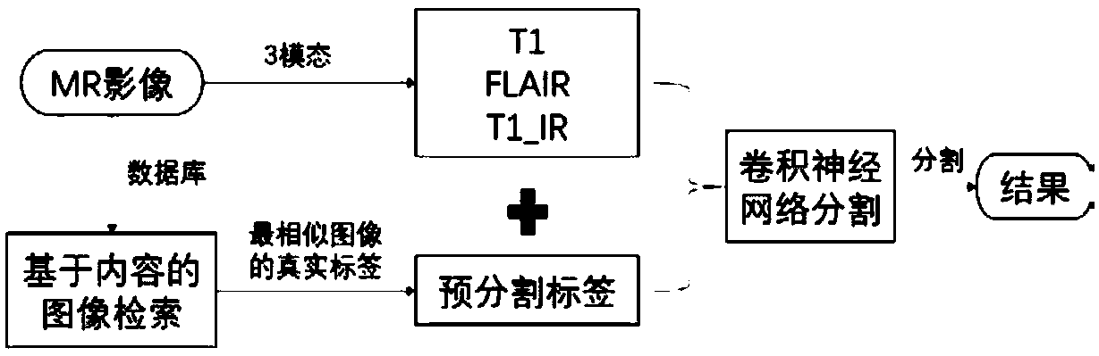
[0073] 1. First, match a set of pre-segmented labels to each set of MRI training data. Since the layer thickness of the data set I used is the same, all the images can be matched only by matching the image on the top of the head. Since the shape of the head is relatively fixed, according to the size of the head, we first locate the top of the head, and then give a range, each time a label is randomly selected within the range as the fourth channel corresponding to another data. In this way, we generate the amount of training data we need to train the network.

[0074] 2. Use the network to train on the training data. Since the input of the network during training is 64*64, we first randomly extract the obtained training data to obtain a series of 64*64*4 training data to train the network.

[0075] 3. Use the trained network to make predictions on the test data.

[0076] First we match the pre-segmented labels to the test data. Since it is a fully convolutional segmentatio...


PUM


Abstract

The invention belongs to the technical field of medical image processing, and particularly relates to a multi-channel head magnetic resonance imaging tissue segmentation method. However, the existingdeep learning method does not utilize anatomical structure information of relatively fixed brain. The invention provides a multi-channel head magnetic resonance imaging tissue segmentation method, which comprises the following steps of: 1, matching a pre-segmentation label most similar to each image to form four channels; And 2, inputting the obtained four channel data into a convolutional neuralnetwork, training the network through a real label of the input data to obtain a training model, and enabling the test data to pass through the trained model to obtain a segmentation result. The priortexture information of brain tissue is fully utilized, a new channel is added, accurate segmentation of the network is promoted, and the network segmentation precision is improved. The method provided by the invention is simple and high in robustness, and can be added to any segmented network for segmentation without changing the original network structure.

Description

technical field [0001] The present application belongs to the technical field of medical image processing, and in particular relates to a multi-channel head magnetic resonance imaging tissue segmentation method. Background technique [0002] Magnetic resonance imaging (magnetic resonance imaging, MRI) is an important modern medical imaging technology. Compared with other medical imaging technologies, MRI has the following advantages: high resolution imaging of soft tissues, no radiation damage to the human body, and Multi-directional imaging. Due to these advantages, MRI is widely used in pathological and functional studies of the brain, such as neurological diseases such as epilepsy, Alzheimer's disease, multiple sclerosis, diagnosis of tumors, and research on the working mechanism of the brain. Moreover, MRI technology is also involved in the treatment of encephalomyopathy, such as radiation therapy for tumors. Accurate segmentation of brain tissue in MRI is the basis fo...

Claims


Application Information

Patent Timeline
IPC IPC(8): G06T7/10
CPCG06T2207/10088G06T2207/20081G06T2207/20084G06T2207/30016G06T7/10
Inventor 袁克虹孙窈张硕
Owner SHENZHEN GRADUATE SCHOOL TSINGHUA UNIV
Why Eureka
Try Eureka