Image quality assessment method and device and computer readable storage medium

An image quality assessment, fundus image technology, applied in the field of medical image processing, to achieve the effect of improving accuracy, ensuring accuracy, and avoiding misdiagnosis and missed diagnosis

Active Publication Date: 2019-04-30
北京康夫子健康技术有限公司
View PDF7 Cites 9 Cited by
  • Summary
  • Abstract
  • Description
  • Claims
  • Application Information

AI Technical Summary

Problems solved by technology

[0003] Embodiments of the present invention provide an image quality assessment method, device

Method used


Image

  • Image quality assessment method and device and computer readable storage medium
  • Image quality assessment method and device and computer readable storage medium
  • Image quality assessment method and device and computer readable storage medium

Examples


Example Embodiment

[0051] In the following, only certain exemplary embodiments are briefly described. As those skilled in the art can realize, the described embodiments may be modified in various different ways without departing from the spirit or scope of the present invention. Therefore, the drawings and description are to be regarded as illustrative in nature and not restrictive.
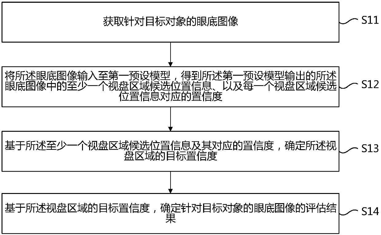
[0052] In one embodiment, figure 1 Shows a flowchart of an image quality evaluation method according to an embodiment of the present invention, the method includes:

[0053] Step S11: Obtain a fundus image for the target object;

[0054] Step S12: Input the fundus image to a first preset model to obtain at least one optic disc region candidate position information corresponding to the optic disc region in the fundus image output by the first preset model, and each optic disc region candidate The confidence level corresponding to the location information;

[0055] Step S13: Determine the target confidence level of the vid...


PUM


Abstract

The embodiment of the invention provides an image quality assessment method and device and a computer readable storage medium. The method comprises the steps that an eye fundus image for a target object is acquired; the fundus image is input into a first preset model to obtain at least one optic disc area candidate position information in the fundus image output by the first preset model and a confidence coefficient corresponding to each optic disc area candidate position information; based on the candidate position information of the at least one optic disk area and the corresponding confidence coefficient, a target confidence coefficient of the optic disk area is determined; and an evaluation result of the fundus image is determined for the target object based on the target confidence coefficient of the optic disc area. The problems that the medical image analysis scene requirement cannot be met and the accuracy of an output result cannot be ensured are solved.

Description

technical field [0001] The present invention relates to the technical field of medical image processing, in particular to an image quality evaluation method, device and computer-readable storage medium. Background technique [0002] Retinal fundus maps play an important role in the screening and diagnosis of fundus diseases. A clear fundus map is a prerequisite for the intelligent fundus screening system to obtain reliable diagnostic results. At present, the schemes generally adopted for fundus image quality assessment include: assessing image quality based on a general image quality control module; and / or performing quality assessment based on extracting statistical features of a local region of interest. However, the main problem of the existing methods is that they cannot meet the requirements of medical image analysis scenarios and cannot guarantee the accuracy of the output results. Contents of the invention [0003] Embodiments of the present invention provide an i...

Claims


Application Information

Patent Timeline
IPC IPC(8): G06T7/00
CPCG06T7/0012G06T2207/30041G06T2207/30168
Inventor 孙旭杨叶辉王磊许言午黄艳
Owner 北京康夫子健康技术有限公司
Why Eureka
Try Eureka