Iterative Update Super-resolution Microscopic Imaging Method of Stripe Illumination in Fourier Domain Based on Total Internal Reflection
What is AI technical title?
AI technical title is built by Patsnap AI team. It summarizes the technical point description of the patent document.
An iterative update, microscopic imaging technology, applied in the field of optical super-resolution microscopic imaging, which can solve problems such as difficult to apply in vivo imaging
Active Publication Date: 2020-10-30
ZHEJIANG UNIV
View PDF7 Cites 0 Cited by
Summary
Abstract
Description
Claims
Application Information
AI Technical Summary
This helps you quickly interpret patents by identifying the three key elements:
Problems solved by technology
Method used
Benefits of technology
Problems solved by technology
Other methods such as Photoactivated Localization Microscopy (PALM) and Stochastic Light Reconstruction Microscopy (STORM) are much slower than SIM for large fields of view, and they are difficult to reconstruct a super-resolution image. , requires thousands of original images, which makes it difficult to apply this method to in vivo imaging
Method used
Image
Click on the blue label to locate the original text in one second.
Reading with bidirectional positioning of images and text.
Examples
Embodiment 1
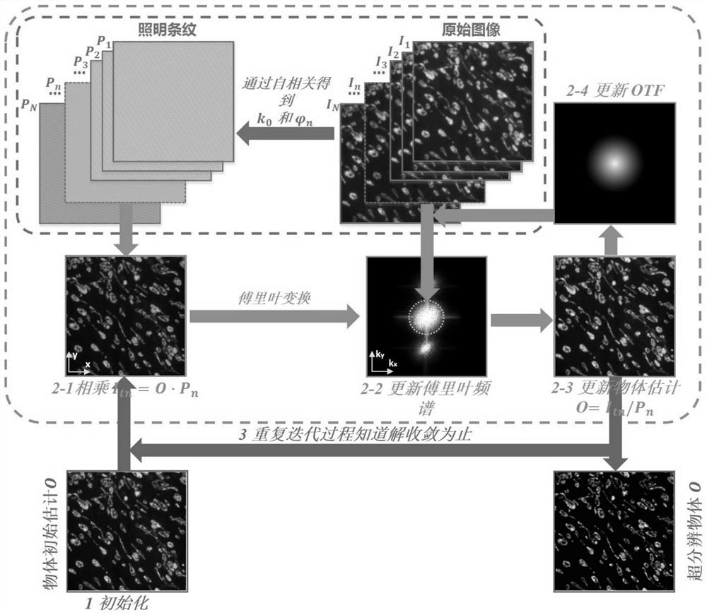
[0059] Such as figure 1 As shown, a method for iteratively updating super-resolution microscopic imaging in the Fourier domain of stripe illumination based on total internal reflection provided in this embodiment includes the following steps:
[0060] (1) Split a beam of parallel illuminated laser beams into two beams of parallel beams with equal intensity and consistent polarization direction, converge to the entrance pupil surface of the objective lens, and then become two beams of parallel beams after passing through the objective lens. The surface excites two counter-propagating evanescent waves for interference, producing fine evanescent wave stripes to illuminate the fluorescent sample, and the fluorescent sample is modulated by the non-uniform illumination light field, and the frequency spectrum shifts; after the fluorescent signal emitted by the fluorescent sample is received by the objective lens , the fluorescent signal is received by the detector on the imaging imag...
Embodiment 2
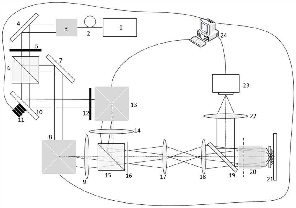
[0102] Such as figure 2 A super-resolution microscopic imaging device for realizing the method of the present invention is provided, but not limited to figure 2 device shown.
[0103] The fringe illumination Fourier domain iterative update super-resolution microscopic imaging device based on total internal reflection in this embodiment includes a laser 1, a polarization-maintaining single-mode fiber 2, a beam collimator 3, a first reflector 4, a first dichotomous A wave plate 5, a polarizing beam splitter 6, a second mirror 7, a first vibration mirror module 8, a first scanning lens 9, a third mirror 10, a piezoelectric ceramic 11, and a second half-wave plate 12. Second galvanometer module 13, second scanning lens 14, beam combiner 15, polarization converter 16, first field lens 17, second field lens 18, dichroic mirror 19, microscope objective lens 20, sample 21 , the third field mirror 22, EMCCD 23, computer 24.
[0104] use figure 2 The wide-field super-resolution m...
PUM
Abstract
The invention discloses a fringe illumination Fourier domain iteration updating super-resolution microscopic imaging method based on total internal reflection. The method comprises the following steps: splitting a parallel illumination laser beam into two parallel beams with equal intensity and consistent polarization direction, and exciting two oppositely propagated evanescent waves to interfereso as to generate a fine evanescent wave fringe illumination fluorescent sample; receiving a fluorescence signal on the imaging image surface by using a detector to obtain a low-resolution image mixedwith high-frequency and low-frequency information of the fluorescence sample; changing the spatial displacement and direction of the evanescent wave illumination fringes for multiple times, and shooting the fluorescence signal modulated by the fringe intensity again to obtain a series of low-resolution images mixed with high-frequency and low-frequency information of the fluorescence sample as original images; and finally, carrying out Fourier domain iteration updating processing on the original image, and carrying out continuous iteration to finally reconstruct a super-resolution image of the fluorescent sample. According to the method, the transverse resolution of about 100 nm can be achieved, the background level can be reduced, the contrast ratio can be improved, the unknown aberration of the system can be corrected, and in-vivo imaging can be achieved.
Description
technical field [0001] The invention relates to the field of optical super-resolution microscopic imaging, in particular to a method for iteratively updating super-resolution microscopic imaging in the Fourier domain of fringe illumination based on total internal reflection. Background technique [0002] Fluorescence microscopy plays a pivotal role in the application of biological research and clinical diagnosis due to its non-invasive characteristics. However, due to the existence of the diffraction limit, the resolution of optical microscopy usually cannot exceed 200nm. In the past few decades, many techniques have been invented to break through this diffraction limit. Among these microscopy techniques, structured illumination microscopy (SIM) is a powerful tool in biomedical imaging because the The method can provide high temporal and spatial resolution, and can achieve video-rate imaging speed. Although SIM can only achieve a two-fold improvement in resolution, it requ...
Claims
Application Information
Patent Timeline
Application Date:The date an application was filed.
Publication Date:The date a patent or application was officially published.
First Publication Date:The earliest publication date of a patent with the same application number.
Issue Date:Publication date of the patent grant document.
PCT Entry Date:The Entry date of PCT National Phase.
Estimated Expiry Date:The statutory expiry date of a patent right according to the Patent Law, and it is the longest term of protection that the patent right can achieve without the termination of the patent right due to other reasons(Term extension factor has been taken into account ).
Invalid Date:Actual expiry date is based on effective date or publication date of legal transaction data of invalid patent.

