A fringe illumination Fourier domain iteration updating super-resolution microscopic imaging method based on total internal reflection

An iterative update and microscopic imaging technology, applied in the field of optical super-resolution microscopic imaging, can solve problems such as difficult application of in vivo imaging

Active Publication Date: 2019-05-03
ZHEJIANG UNIV
View PDF7 Cites 11 Cited by
  • Summary
  • Abstract
  • Description
  • Claims
  • Application Information

AI Technical Summary

Problems solved by technology

Other methods such as Photoactivated Localization Microscopy (PALM) and Stochastic Light Reconstruction Microscopy (STORM) are much slower than SIM for large fields of view, and they are difficult to reconstruct a super-resolution image. , requires thousands of original images, which makes it difficult to apply this method to in vivo imaging

Method used


Image

  • A fringe illumination Fourier domain iteration updating super-resolution microscopic imaging method based on total internal reflection
  • A fringe illumination Fourier domain iteration updating super-resolution microscopic imaging method based on total internal reflection
  • A fringe illumination Fourier domain iteration updating super-resolution microscopic imaging method based on total internal reflection

Examples


Embodiment 1

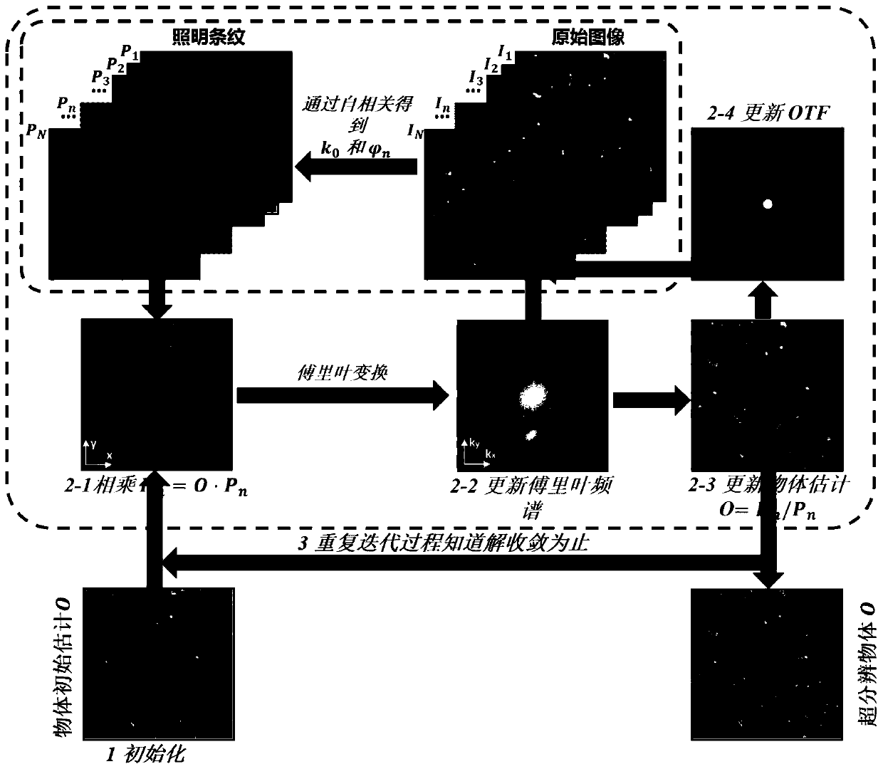
[0059] Such as figure 1 As shown, this embodiment provides a fringe-illuminated Fourier-domain iterative update super-resolution microscopy imaging method based on total internal reflection, including the following steps:

[0060] (1) Split a parallel-illuminated laser beam into two parallel beams with equal intensity and the same polarization direction, converge on the entrance pupil surface of the objective lens, and then pass through the objective lens to become two parallel beams. The surface excites two evanescent waves propagating in opposite directions to interfere, and produce fine evanescent wave fringes to illuminate the fluorescent sample. After the fluorescent sample is modulated by the non-uniform illumination light field, the spectrum shifts; after receiving the fluorescent signal from the fluorescent sample by the objective lens , Use a detector to receive the fluorescence signal on the imaging image plane to obtain a low resolution image mixed with high and low fr...

Embodiment 2

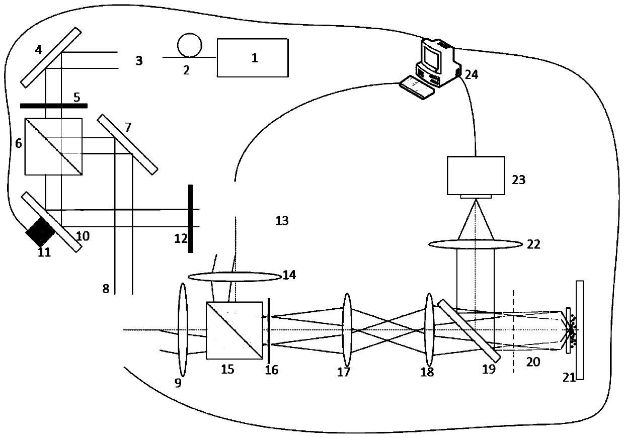
[0102] Such as figure 2 A super-resolution microscopic imaging device for implementing the method of the present invention is given, but not limited to figure 2 The device shown.

[0103] The fringe illumination Fourier domain iteratively updated super-resolution microscopic imaging device based on total internal reflection in this embodiment includes a laser 1, a polarization-maintaining single-mode fiber 2, a beam collimator 3, a first mirror 4, and a first bisection One-wave plate 5, polarization beam splitter 6, second mirror 7, first galvanometer module 8, first scanning lens 9, third mirror 10, piezoelectric ceramic 11, second half-wave plate 12. The second galvanometer module 13, the second scanning lens 14, the beam combiner 15, the polarization converter 16, the first field lens 17, the second field lens 18, the dichroic lens 19, the microscope objective lens 20, the sample 21 , The third field lens 22, EMCCD 23, computer 24.

[0104] use figure 2 The process of wide-f...


PUM


Abstract

The invention discloses a fringe illumination Fourier domain iteration updating super-resolution microscopic imaging method based on total internal reflection. The method comprises the following steps: splitting a parallel illumination laser beam into two parallel beams with equal intensity and consistent polarization direction, and exciting two oppositely propagated evanescent waves to interfereso as to generate a fine evanescent wave fringe illumination fluorescent sample; receiving a fluorescence signal on the imaging image surface by using a detector to obtain a low-resolution image mixedwith high-frequency and low-frequency information of the fluorescence sample; changing the spatial displacement and direction of the evanescent wave illumination fringes for multiple times, and shooting the fluorescence signal modulated by the fringe intensity again to obtain a series of low-resolution images mixed with high-frequency and low-frequency information of the fluorescence sample as original images; and finally, carrying out Fourier domain iteration updating processing on the original image, and carrying out continuous iteration to finally reconstruct a super-resolution image of the fluorescent sample. According to the method, the transverse resolution of about 100 nm can be achieved, the background level can be reduced, the contrast ratio can be improved, the unknown aberration of the system can be corrected, and in-vivo imaging can be achieved.

Description

Technical field [0001] The invention relates to the field of optical super-resolution microscopic imaging, in particular to a fringe illumination Fourier domain iterative update super-resolution microscopic imaging method based on total internal reflection. Background technique [0002] Fluorescence microscopy plays an important role in the application of biological research and diagnosis in Linchuan because of its non-invasive characteristics. However, due to the diffraction limit, the resolution of optical microscopes usually cannot exceed 200 nm. In the past few decades, people have invented many technologies to break this diffraction limit. Among these microscopy, structured illumination microscopy (SIM) is a powerful tool in biomedical imaging because of the The method can provide higher temporal and spatial resolution, and can achieve imaging speed of video rate. Although SIM can only achieve a two-fold increase in resolution, the optical power it requires is much smaller...

Claims


Application Information

Patent Timeline
Patent Type & Authority Applications(China)
IPC IPC(8): G06T3/40G01N21/64G02B21/36
Inventor 刘旭刘秋兰匡翠方刘文杰
Owner ZHEJIANG UNIV
Why Eureka
Try Eureka