A three-dimensional tumor contour reconstruction method for focus observation and diagnosis through an HIFU system

A technology of 3D reconstruction and 3D contour, applied in 3D image processing, image data processing, instruments, etc., can solve the problems of inability to automatically treat tumors with conformal shape, and help analyze the treatment effect, facilitate and accurately outline, and ensure safety and effectiveness Effect

Active Publication Date: 2019-05-07
HAIYING ELECTRONIC MEDICAL SYST CO LTD
View PDF2 Cites 1 Cited by
  • Summary
  • Abstract
  • Description
  • Claims
  • Application Information

AI Technical Summary

Problems solved by technology

[0004] In the existing treatment methods, the operator cannot fully understand the entire tumor shape in the HIFU treatment system during the treatment process, and rationally plan the treatment and control plan for the overall tumor body, so that it is impossible to perform conformal automatic treatment on the entire tumor body, and manually adjust points in parallel. Front spacing, re-circle, etc.

Method used


Image

  • A three-dimensional tumor contour reconstruction method for focus observation and diagnosis through an HIFU system
  • A three-dimensional tumor contour reconstruction method for focus observation and diagnosis through an HIFU system
  • A three-dimensional tumor contour reconstruction method for focus observation and diagnosis through an HIFU system

Examples


Embodiment 1

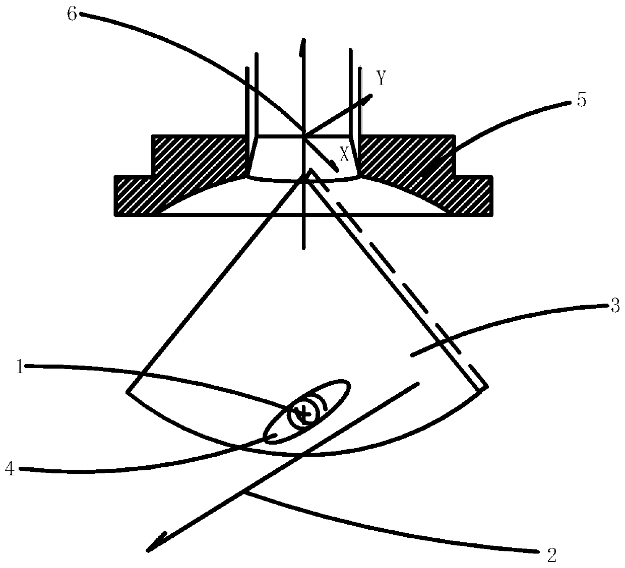
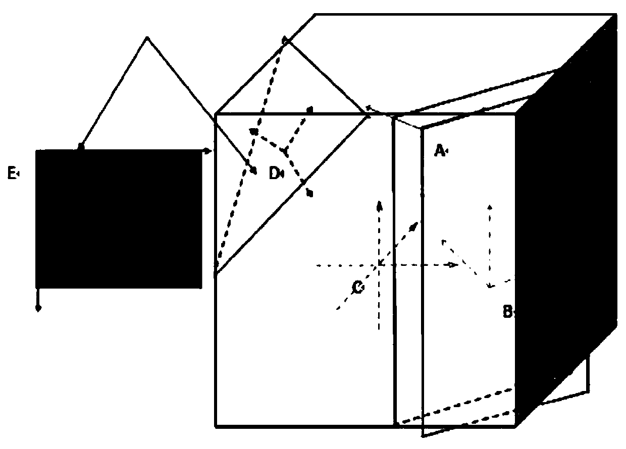
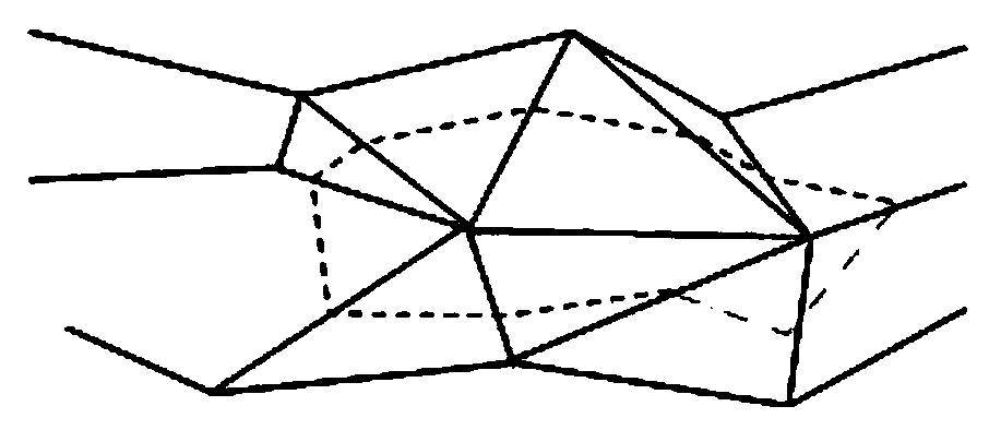
[0045] See Figure 1 to Figure 3 , the present invention specifically comprises the following steps:

[0046] a. Accurate B-ultrasound image scanning of the tumor tissue through the positioning B-ultrasound probe installed in the central hole directly above the treatment transducer in the HIFU treatment system;

[0047] b. For a group of B-ultrasound two-dimensional images collected by the HIFU treatment system, manually outline the tumor area and sensitive area on the background of the two-dimensional B-ultrasound image;

[0048] c. The HIFU treatment system constructs a three-dimensional contour body after outlining. According to a series of B-ultrasound images scanned precisely and the depth of the coronal plane, linear difference calculation is performed to obtain image information of the coronal plane and generate visualized coronal planes of different depths. face image;

[0049] d. According to the three-dimensional contour and cut plane, select the angle, plan the cu...


PUM


Abstract

The invention relates to a three-dimensional tumor contour reconstruction method for focus observation and diagnosis through an HIFU system. The three-dimensional tumor contour reconstruction method comprises the following steps: carrying out image scanning on a tumor tissue part through a positioning B ultrasonic probe; Carrying out manual edge hooking on a tumor region and a sensitive region onthe background of the acquired two-dimensional B-ultrasonic image; enabling The constructed three-dimensional contour body to carry out linear difference calculation according to a series of preciselyscanned B-ultrasonic images and the depth of the coronal plane, obtaining image information of the coronal plane, and generating visual coronal plane images with different depths; According to the three-dimensional contour body and the tangent plane, selecting an angle, and planning and treating the tangent plane; Collecting the B-mode ultrasound image again according to the original parameters,manually carrying out edge hooking on an echo enhancement area of the B-mode ultrasound image, and constructing a static three-dimensional contour body image after treatment; And constructing a three-dimensional tumor data storage format. According to the invention, the tumor contour is marked on the two-dimensional image, and then the three-dimensional tumor shape is established. The tangent plane boundary contour in any direction is the intersection line of the tangent plane and the three-dimensional tumor.

Description

technical field [0001] The invention relates to the technical field of HIFU treatment systems, in particular to a method for three-dimensional reconstruction of tumor contours for HIFU system observation and diagnosis of lesions. Background technique [0002] HIFU stands for High Intensity Focused Ultrasound Therapeutic System, which utilizes the beam radiation and good penetrating power of ultrasound in the low megahertz frequency band to focus the ultrasound energy in the desired treatment tissue of the human body in a certain focusing way to form an acoustic Higher intensity area—the focal region. The tissue in the focal region absorbs ultrasonic energy and converts it into heat energy to increase its temperature. When the sound intensity in the focal region reaches thousands or even W / cm2, the temperature will be between 0.5 and It rises to above 70°C within 5s, leading to coagulation necrosis of tissue cells—that is, ablation, and loss of proliferation, infiltration and...

Claims


Application Information

Patent Timeline
Patent Type & Authority Applications(China)
IPC IPC(8): G06T7/00G06T15/00
Inventor 王晓逸耿晓鸣王国英
Owner HAIYING ELECTRONIC MEDICAL SYST CO LTD
Why Eureka
Try Eureka