
Computed tomography device

A technology of tomography and computer, applied in computer tomography scanner, diagnosis, echo tomography, etc., can solve the problem that doctors cannot make treatment decisions

Pending Publication Date: 2019-07-16
SIEMENS HEALTHINEERS LTD
View PDF8 Cites 0 Cited by
  • Summary
  • Abstract
  • Description
  • Claims
  • Application Information

AI Technical Summary

Problems solved by technology

However, clinically, physicians cannot make rapid treatment decisions based on these parameters alone

Method used


Image

  • Computed tomography device

Examples


Embodiment Construction

[0026] In order to make the purpose, technical solution and advantages of the present invention clearer, the following examples are given to further describe the present invention in detail.

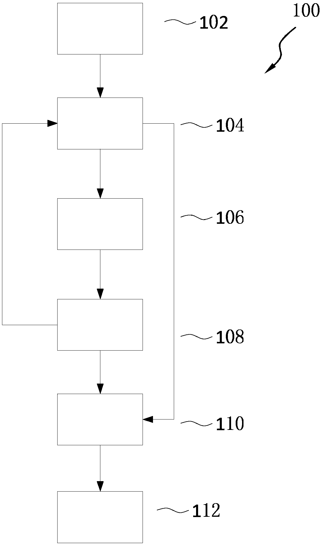
[0027] figure 1 is a structural block diagram of the computed tomography device 100 according to an embodiment of the present invention. Such as figure 1 As shown, the computed tomography device 100 includes a storage unit 104 , a scanning unit 106 , a measuring unit 108 and a comparison unit 110 . The computed tomography device 100 can be used to evaluate the treatment effect of a certain subject during the time period from the first time to the second time, for example, the effect of treating a tumor, wherein the first time is earlier than the second time.

[0028] The storage unit 104 stores scan parameters and contrast agent injection parameters. Scan parameters may include tube voltage, tube current, scan range and scan phase. Contrast injection parameters may include injection ...


PUM


Abstract

The present invention discloses a computed tomography device. The computed tomography device comprises a storage unit, a scanning unit, a measurement unit and a comparison unit; the storage unit is used for storing scan parameters and contrast injection parameters; the scanning unit respectively acquires a first set of dual energy tomographic images and a second set of dual energy tomographic images by using the scan parameters and contrast agent injection parameters at a first time and a second time respectively; the measurement unit measures quantitative parameters of lesions in the first set of the dual energy tomographic images and the quantitative parameters of the same lesions in the second set of the dual energy tomographic images; and the comparison unit compares significant differences between the quantitative parameters of the lesions in the first set of the dual energy tomographic images and the quantitative parameters of the lesions in the second set of dual energy tomographic images. The computed tomography device acquires the dual-energy tomographic images of multiple time nodes using the same personalized scan parameters and contrast agent injection parameters for anentire course of a same patient, measures the quantitative parameters of the lesions, and determines the significant differences therefrom.

Description

technical field [0001] The present invention relates to computed tomography, in particular dual energy computed tomography. Background technique [0002] Contrast-enhanced CT is widely used in tumor therapy assessment. However, different patients with different types of tumors will have different time attenuation curves, resulting in different scan phases with the best image quality as well as different optimal contrast agent doses and minimum radiation doses. Conventional scanning protocols cannot provide personalized data acquisition and precise quantitative measurement, thus may generate false tumor response assessment reports and lead to wrong decisions on subsequent treatment. [0003] Currently, most measurements of tumor changes after treatment are manual and qualitative. Especially for some tumors of hollow organs, such as gastric cancer, colorectal cancer, etc., the measurement is unstable and unrepeatable. In addition, with the development of CT technology and b...

Claims


Application Information

Patent Timeline
Patent Type & Authority Applications(China)
IPC IPC(8): A61B6/03
CPCA61B6/032A61B6/4411A61B6/481A61B6/5217
Inventor 闫静
Owner SIEMENS HEALTHINEERS LTD
Learn More