A large joint tissue segmentation method and segmentation system

A tissue and joint technology, applied in the field of medical image processing, can solve problems such as low joint tissue segmentation efficiency, and achieve the effects of simplifying the tissue segmentation process, improving the recognition accuracy, and improving the recognition efficiency.

Active Publication Date: 2020-12-11
北京灵医灵科技有限公司
View PDF12 Cites 0 Cited by
  • Summary
  • Abstract
  • Description
  • Claims
  • Application Information

AI Technical Summary

Problems solved by technology

[0012] In view of the above problems, embodiments of the present invention provide a large joint tissue segmentation method and segmentation system to solve the technical problem of low efficiency in existing joint tissue segmentation

Method used


Image

  • A large joint tissue segmentation method and segmentation system
  • A large joint tissue segmentation method and segmentation system
  • A large joint tissue segmentation method and segmentation system

Examples


Embodiment Construction

[0053] In order to make the purpose, technical solution and advantages of the present invention clearer and clearer, the present invention will be further described below in conjunction with the accompanying drawings and specific embodiments. Apparently, the described embodiments are only some of the embodiments of the present invention, but not all of them. Based on the embodiments of the present invention, all other embodiments obtained by persons of ordinary skill in the art without creative efforts fall within the protection scope of the present invention.

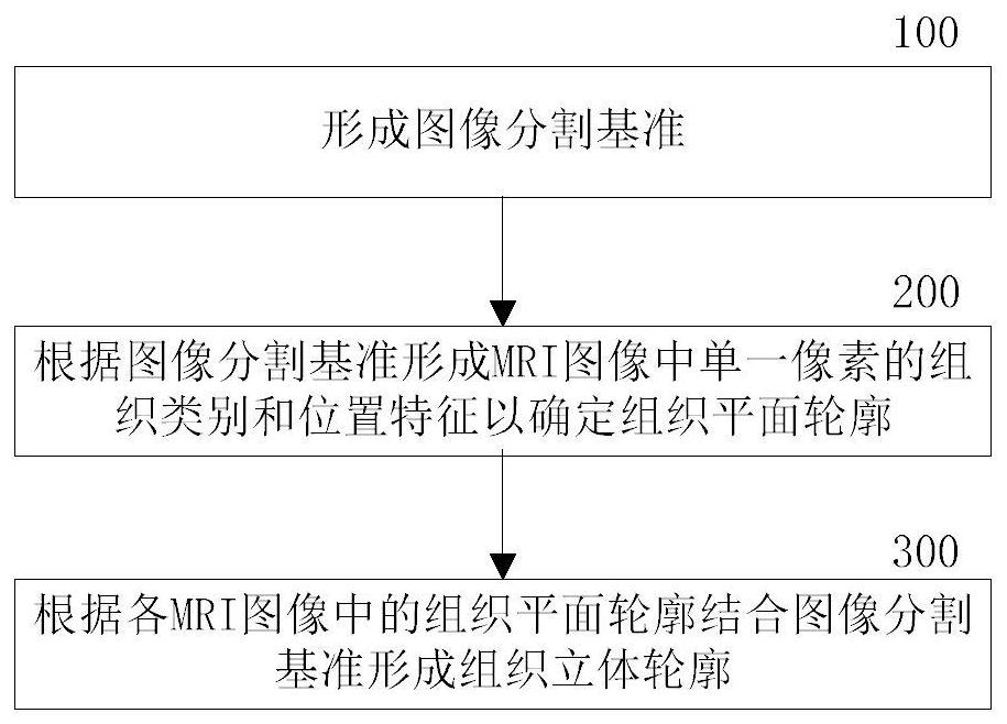
[0054] The segmentation method of large joint tissue in one embodiment of the present invention is as follows: figure 1 shown. exist figure 1 , this example includes:

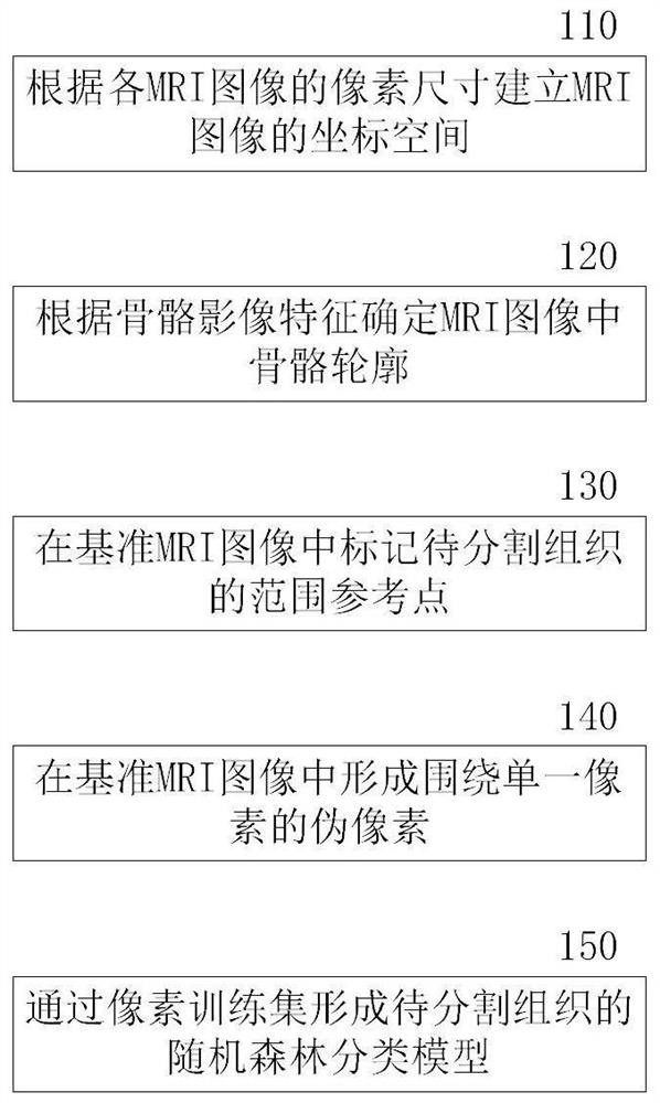
[0055] Step 100: Form an image segmentation benchmark.

[0056]Those skilled in the art can understand that a set of image references of parallel sections includes but not limited to a plane coordinate reference within an image, a coordinate transfo...


PUM


Abstract

The invention provides a large joint tissue segmentation method and a segmentation system, which solves the technical problem of low efficiency of joint tissue segmentation in the prior art. The method includes: forming an image segmentation criterion; forming the tissue category and position feature of a single pixel in the MRI image according to the image segmentation criterion to determine the tissue plane profile; combining the image segmentation with the tissue plane profile in each of the MRI images The fiducials form a three-dimensional outline of the tissue. The use of bone features simplifies the tissue segmentation process, the use of related image and information features to form automatic processing of object contours and the improvement of recognition accuracy, and the use of segmentation benchmarks to form image quantification rules, pixel connotation information, and modeling frameworks. Organizations can achieve automatic subject segmentation, accurate contour positioning, and automatic 3D modeling in MRI images, effectively improving the identification efficiency of professional human identification resources.

Description

technical field [0001] The invention relates to the technical field of medical image processing, in particular to a large joint tissue segmentation method and segmentation system. Background technique [0002] The composition of joints is complex. Taking the wrist joint as an example, it includes bones, tendons, vessels, and nerves. The contours of tissues other than bones are often unclear due to the gray resolution of MRI (Magnetic Resonance Imaging, MRI). The combination of edge gray areas between tissues leads to difficulties in image segmentation between tissues. [0003] In the prior art, it is necessary to manually perform selective editing, defect compensation processing, artifacts and redundant data separation on various tissue patterns, and then use the region growing method to generate segmentation results to establish a complete digital model, such as large Contour segmentation of bone tissue. This will consume a lot of time for operators, and professional reso...

Claims


Application Information

Patent Timeline
Patent Type & Authority Patents(China)
IPC IPC(8): G06T7/11G06K9/62G06T7/136
CPCG06T7/11G06T7/136G06T2207/10012G06T2207/30008G06F18/2415
Inventor 林海晓武正强
Owner 北京灵医灵科技有限公司
Why Eureka
Try Eureka