MRI automatic image segmentation method based on lesion volume measurement

An automatic image and volume measurement technology, applied in the field of image processing, can solve the problems of not being suitable for large-scale image segmentation operations, affecting the accuracy of case judgment, hindering popularization and promotion, etc., achieving small error, high segmentation efficiency, and high accuracy Effect

Inactive Publication Date: 2017-11-10
ZHEJIANG GONGSHANG UNIVERSITY
View PDF4 Cites 0 Cited by
  • Summary
  • Abstract
  • Description
  • Claims
  • Application Information

AI Technical Summary

Problems solved by technology

The above two methods have their own defects. The defects of the first method are as follows: 1) because the lesion is manually delineated, this method is mostly used in more complicated occasions; It is not suitable for most operators who lack relevant professional knowledge; 3) Since this method uses manual delineation of lesions, the segmentation efficiency is very low, and it is not suitable for large-scale image segmentation operations, which seriously hinders the popularization and promotion of this method; The defects of the two methods are as follows: since the signal intensity of non-brain tissues such as skull, eyeball and muscle and background noise in the MRI scanning image overlaps with the signal intensity of brain tissue to a certain extent, the dynamic fuzzy K-means clustering algorithm is used to segment There will be misclassification, and misclassification will directly affect the accuracy of case judgment. Misjudgment is unacceptable to patients and their families, and it is also the source of most doctor-patient conflicts.

Method used


Image

  • MRI automatic image segmentation method based on lesion volume measurement
  • MRI automatic image segmentation method based on lesion volume measurement
  • MRI automatic image segmentation method based on lesion volume measurement

Examples


Embodiment Construction

[0029] The present invention will be further described in detail below in conjunction with the accompanying drawings and embodiments.

[0030] A kind of MRI automatic image segmentation method based on lesion volume measurement proposed by the present invention, its overall realization block diagram is as follows figure 1 shown, which includes the following steps:

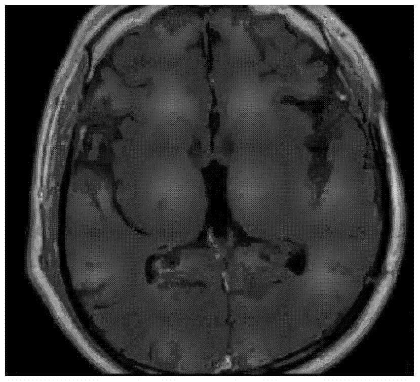
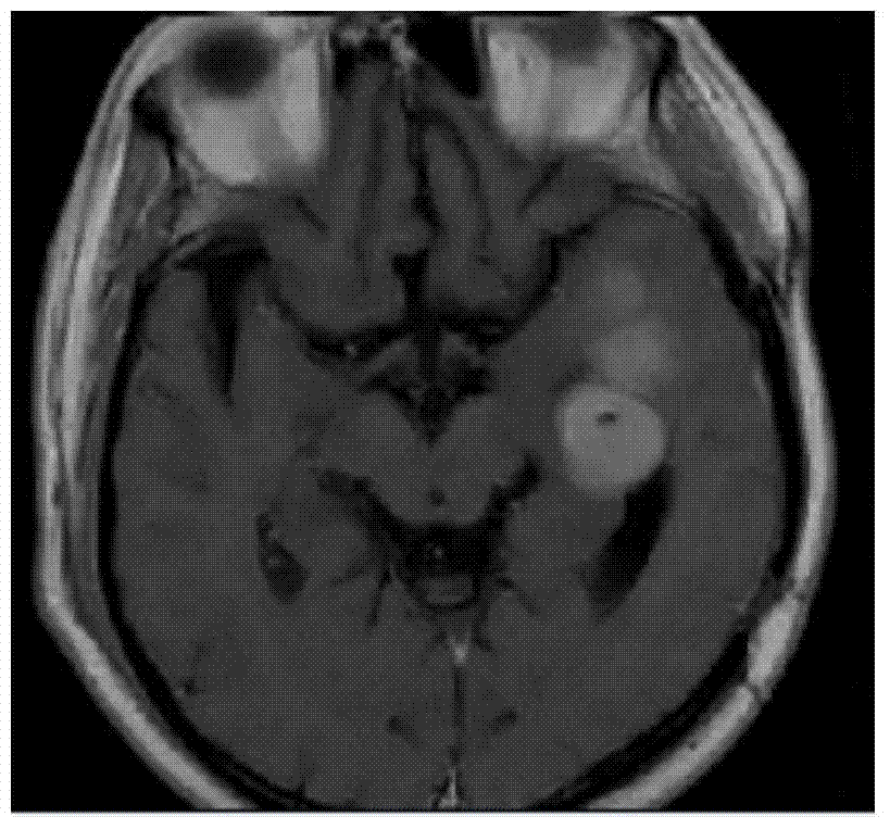
[0031] ① Obtain an MRI scan image from the hospital's MRI medical imaging equipment as the MRI scan image to be segmented, and then convert the MRI scan image to be segmented into a grayscale image.

[0032] ②Assuming that the width and height of the grayscale image correspond to W×H, then if W×H can be divisible by u×u, then define the grayscale image as the current grayscale image, and then directly divide the current grayscale image into non-overlapping sub-blocks of size u×u; if W×H cannot be divisible by u×u, then expand the grayscale image so that its size can be divisible by u×u, and the expanded grayscale...


PUM


Abstract

The present invention discloses a lesion volume measurement based MRI (Magnetic Resonance Imaging) automatic image segmentation method. The method comprises: firstly, acquiring a gray image of a to-be-segmented MRI scanned image; secondly, dividing the gray image into sub-blocks that do not overlap mutually; thirdly, segmenting the gray image by using a marker-based watershed segmentation algorithm to obtain a plurality of preliminary lesion regions; and finally, inputting respective pixel values, as input parameters, of all pixel points in each sub-block corresponding to each preliminary lesion region into a non-linear optimization model for optimization to obtain a corresponding final lesion region. The advantage is that preliminary segmentation is performed on the gray image by using the marker-based watershed segmentation algorithm, so that not only does the obtained preliminary lesion region have high accuracy, but also the segmentation efficiency is high; after the preliminary segmentation, the optimization is performed by using the non-linear optimization model, so that an accurate lesion region can be obtained only by performing segmentation once in combination with optimization; and a segmentation process is simple, the segmentation efficiency is high, and the computation amount is small, so that the method is suitable for massive image segmentation operations.

Description

technical field [0001] The present invention relates to an image processing technology, in particular to an MRI (Magnetic Resonance Imaging, magnetic resonance imaging) automatic image segmentation method based on lesion volume measurement. Background technique [0002] Multiple sclerosis is a demyelinating disease of the central nervous system. It is more common in North America and Europe. It is estimated that there are at least 3 to 4 million patients in the world, accounting for 6% to 10% of the incidence of nervous system diseases. In recent years, reports from Japan and China Increasingly. [0003] Magnetic resonance imaging is the only effective imaging evaluation method for clinical diagnosis and treatment of multiple sclerosis. At present, for experienced radiologists to determine gray and white matter lesions in MRI scan images, the following two methods are usually used. The first method is to manually delineate the region of interest (ROI, region of interest) of...

Claims


Application Information

Patent Timeline
Patent Type & Authority Patents(China)
IPC IPC(8): G06T7/00G06T7/11
CPCG06T7/0012G06T2207/10088G06T2207/30016
Inventor 傅均汤旭翔赵帅陈赛陈柳柳曹海洋
Owner ZHEJIANG GONGSHANG UNIVERSITY
Why Eureka
Try Eureka