Method for interactively segmenting lung lobes

An interactive, lobe technology, applied in the field of medical image processing, can solve the problems of lack of explanation of key steps and low accuracy, and achieve the effect of convenient diagnosis and treatment

Active Publication Date: 2019-11-19
SAINUO WEISHENG SCI & TECH BEIJING
View PDF4 Cites 8 Cited by
  • Summary
  • Abstract
  • Description
  • Claims
  • Application Information

AI Technical Summary

Problems solved by technology

However, the key steps of how to obtain the real lung lobe from the position of the lobe fissure are not explained
In addition, the accuracy of this method is low, and it takes a long time to fit the surface

Method used


Image

  • Method for interactively segmenting lung lobes
  • Method for interactively segmenting lung lobes
  • Method for interactively segmenting lung lobes

Examples


Embodiment Construction

[0041] The present invention will be described in detail below in conjunction with the accompanying drawings and embodiments. It should be noted that various modifications can be made to the embodiments disclosed herein, therefore, the embodiments disclosed in the specification should not be regarded as limitations on the present invention, but only as examples of embodiments, and its purpose is to make the present invention The features of the invention are self-evident.

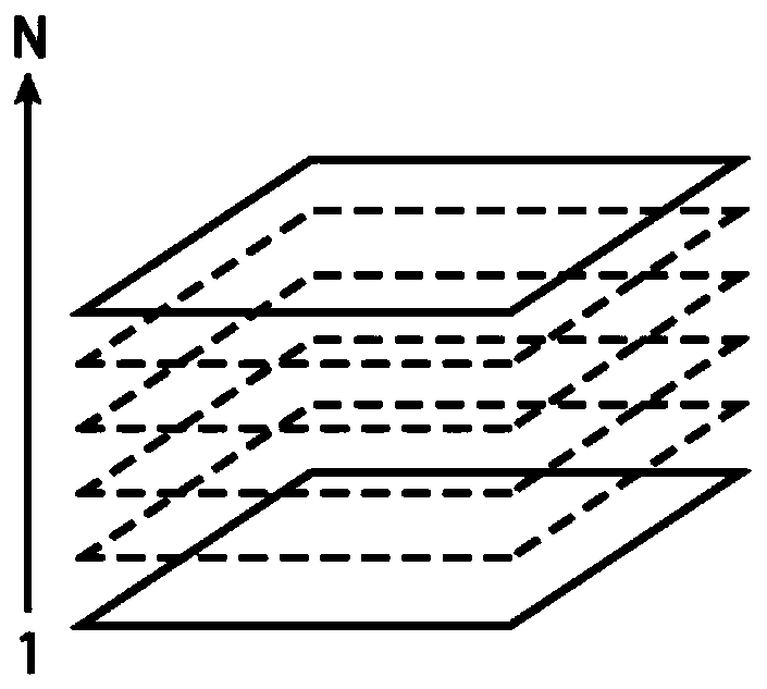
[0042] The structure of human lungs is complex. It is divided into left lung and right lung. The left lung consists of two leaves, the left upper lobe and the left lower lobe. The right lung consists of three leaves, namely the right upper lobe, right middle lobe and right lower lobe. The leaves are covered with Intricate pulmonary trachea and pulmonary vessels. When a CT machine scans the human lungs, such as figure 1 As shown, the output scan data is output layer by layer from bottom to top, that is, t...


PUM


Abstract

The invention provides a method for interactively segmenting lung lobes. The method comprises the following steps: A, automatically separating a left lung from a right lung; B, interactively segmenting lung lobes of the left lung and the right lung, namely S1, interactively describing lung crack tracks; S2, finding all lung fracture points connected with the known lung fracture points on the finding plane according to the points of the known lung fractures, the similarity of the lung fractures and a similarity energy minimization method; S3, growing an upper region and a lower region accordingto the lung fracture points on the searching plane; if the lung is the left lung, directly taking a lung crack as a boundary, and segmenting two parts, namely an upper leaf and a lower leaf, of the left lung; if the lung is the right lung, segmenting the upper middle part and the lower right leaf of the right lung by taking the lung crack as a boundary, then executing the steps S1 to S3 repeatedly in the upper middle part of the right lung, and segmenting the upper middle part of the right lung into the upper right lung leaf and the middle right lung leaf according to the new lung crack on the finding plane.

Description

technical field [0001] The present invention relates to a method for interactively segmenting lung lobes. The invention belongs to the technical field of medical image processing. Background technique [0002] Usually, clinicians diagnose diseases by reading CT (Computed Tomography) images, so as to propose possible treatment options. However, CT images are three-dimensional tomographic images. A chest CT image containing the entire lung usually contains hundreds of two-dimensional slice images. It becomes more, usually there are more than 500 two-dimensional slice images of HRCT of a complete lung. [0003] If clinicians want to accurately diagnose the diseases that may exist in the same thoracic cavity, they need to interpret more than 500 two-dimensional CT slice images, which is a huge workload! This kind of heavy work is not only time-consuming, but also easy to make people feel tired, which may lead to misjudgment and missed diagnosis of diseases. [0004] In this ...

Claims


Application Information

Patent Timeline
Patent Type & Authority Applications(China)
IPC IPC(8): G06T7/11G06T7/187G06T7/136G16H30/20
CPCG06T7/11G06T7/187G06T7/136G16H30/20G06T2207/10081G06T2207/20004G06T2207/30061
Inventor 马双
Owner SAINUO WEISHENG SCI & TECH BEIJING
Why Eureka
Try Eureka