Water-soluble fluorescence probe and nanoparticles with aggregation induction emission effect for ovarian cancer as well as preparation method and application thereof
A technology of aggregation-induced emission and fluorescent molecular probes, applied in the fields of biological analysis and detection, and chemical analysis, can solve the problems of time and space instability, and achieve the effects of low cost, simple preparation process and short response time.
- Summary
- Abstract
- Description
- Claims
- Application Information
AI Technical Summary
Problems solved by technology
Method used
Image

Examples
Embodiment 1
[0054] The synthesis of compound 3 with enhanced aggregation-induced fluorescence emission of the present invention, the synthetic route is as follows:
[0055]
[0056] Synthesis of compound 1: 4-formylpyridine (107.1 mg, 1 mmol) and aniline (93.1 mg, 1 mmol) were weighed and dissolved in 6-8 mL of glacial acetic acid, and stirred at room temperature for 1 hour. Anisole (207.2 mg, 1 mmol) and ammonium acetate (462.5 mg, 6 mmol) were sequentially added to the reaction system, reacted overnight at 120 °C, quenched the reaction with water, poured the reaction system into 200 mL of ice water, The pH of the system was adjusted to neutral with 0.1 mmol / L sodium hydroxide solution, the mixture was filtered and washed three times with water, dried in vacuum and purified by silica gel column chromatography to obtain the product. The yield was 21.9%. The NMR analysis results are: 1 HNMR (500 MHz, CDCl 3 ) δ 8.47 (d,2H), 7.55 (d,2H), 7.45–7.25 (m, 5H), 7.09 (t,2H), 7.05 (d,2H), 6....
Embodiment 2
[0060] Preparation of water-soluble organic fluorescent molecular probe nanoparticles:
[0061] Use a pipette gun to take 40 µL of compound 3 (1 mM) phosphate buffer solution into the ep tube, and then add 1960 µL of phosphate buffer solution (pH=7.4) under ultrasonic conditions to make the final concentration of compound 3 to 20 µM. The system was stirred at room temperature for 30 minutes to produce opalescence. In order to verify its nano-aggregation behavior, the average particle size was determined to be 600 nm by dynamic light scattering experiments, as shown in the attached figure 1 shown.
Embodiment 3
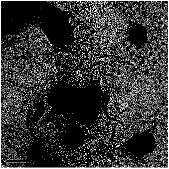
[0063] Scanning electron microscope (SEM) test for the recognition of prepared fluorescent molecular probe nanoparticles and lysophosphatidic acid:
[0064]Take 40 µL of the phosphate buffer solution of compound 3 (1 mM) into the ep tube with a pipette gun, add 1900 µL of the phosphate buffer solution (pH=7.4) under ultrasonic conditions, and then add 60 µL of the phosphate buffer solution of lysophosphatidic acid (1 mM) so that the final concentration of compound 3 in the solution was 20 µM, the final concentration of lysophosphatidic acid was 30 µM, and stirred at room temperature for 30 min. Take a drop of the prepared solution, drop it on the copper grid, blot the water with filter paper, dry it naturally, and put it in a transmission electron microscope for observation. Transmission electron microscope pictures are attached figure 2 shown.
PUM
| Property | Measurement | Unit |
|---|---|---|
| particle size | aaaaa | aaaaa |
| wavelength | aaaaa | aaaaa |
Abstract
Description
Claims
Application Information
 - R&D Engineer
- R&D Manager
- IP Professional
- Industry Leading Data Capabilities
- Powerful AI technology
- Patent DNA Extraction
Browse by: Latest US Patents, China's latest patents, Technical Efficacy Thesaurus, Application Domain, Technology Topic.
© 2024 PatSnap. All rights reserved.Legal|Privacy policy|Modern Slavery Act Transparency Statement|Sitemap