Pathological section interpretation method and system

A pathological sectioning and slicing technology, applied in the field of pathological diagnosis, to achieve the effect of fast and accurate treatment, convenience, accuracy, and cost-free

Inactive Publication Date: 2020-02-07
杭州迪英加科技有限公司
View PDF9 Cites 14 Cited by
  • Summary
  • Abstract
  • Description
  • Claims
  • Application Information

AI Technical Summary

Problems solved by technology

[0003] The purpose of the present invention is to solve the problem in the prior art that the entity PD-L1 immunohistochemical staining section needs to be scanned into a full-field image un

Method used


Image

  • Pathological section interpretation method and system
  • Pathological section interpretation method and system
  • Pathological section interpretation method and system

Examples


Embodiment Construction

[0021] The present invention will be further described below in conjunction with accompanying drawing:
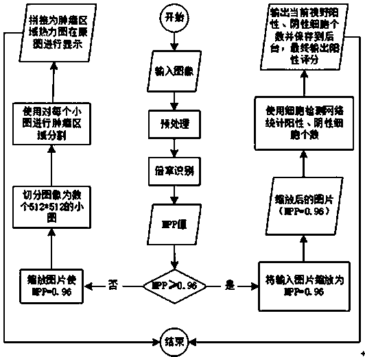
[0022] The MPP values ​​of the digital pathological full-field images used in this embodiment are 0.48, 0.96, 1.92, and 3.84 respectively. The MPP values ​​of images collected by scanners produced by different manufacturers are slightly different at the same magnification, but it does not affect The accuracy of the diagnosis results using the method provided in this application. The microscope used is an in-line microscope, including 1 eyepiece and 4 objective lenses; the magnification of the eyepiece is 10 times, the magnification of the objective lens is 4 times, 10 times, 20 times, 40 times, the images collected by the microscope camera The MPP values ​​are 1.5, 0.6, 0.3 and 0.15, respectively.

[0023] A pathological slice interpretation system, comprising: a microscope, a microscope camera, a processor and a display. Microscope for viewing solid PD-L1 immunohistochem...


PUM


Abstract

The invention relates to a pathological section interpretation method, which is suitable for a pathological section interpretation system. The method comprises the following steps that S10, a microscope camera collects a pathological image of a solid PD-L1 immunohistochemical staining section under a microscope, and sends the pathological image to a processor; S20, the processor preprocesses the pathological image, so that the HSV color space of the pathological image is consistent with a set threshold value; S30, the processor processes, detects and marks the preprocessed pathological image,and outputs a marked pathological image; S40, the display receives and displays the marked pathological image from the processor. The invention further discloses a pathological section interpretationsystem. A computer-readable storage medium stores a computer program for use in conjunction with a pathological section interpretation system. According to the application, the artificial intelligencemethod is used to improve the diagnosis efficiency of doctors and accurately and quickly provide PD-L1 positive scores to assist doctors in clinical judgment.

Description

technical field [0001] The invention relates to the field of pathological diagnosis, in particular to a method and system for interpreting pathological slices. Background technique [0002] Cancer is a threat to human health. In recent years, PD-L1-targeted drugs are expected to become a new method of cancer treatment. The full name of PD-1 is "programmed cell death protein 1", which is an important immunosuppressive molecule. PD-1 immunotherapy is a very representative treatment for tumors. PD-L1 and PD-1 are part of the immune check signaling pathway, which can inhibit the proliferation of T cells and immune defense against tumor cells. PD-L1 and PD-1 work together to inhibit the T cell receptor signaling pathway. PD-L1 targeting drugs bind to PD-L1 to block the combination of PD-L1 and PD-1 on activated T cells, thereby making T cells Still in an activated state to kill tumor cells. PD-1 is mainly expressed on the surface of activated T lymphocytes, B lymphocytes and ...

Claims


Application Information

Patent Timeline
IPC IPC(8): G01N21/84G01N33/574G01N33/68G02B21/36G06K9/62
CPCG01N21/84G01N33/57484G01N33/6803G02B21/365G06F18/241
Inventor 李涵生崔灿慧文丽杨林崔磊
Owner 杭州迪英加科技有限公司
Why Eureka
Try Eureka