Eye fundus image optic disc and macular positioning detection algorithm based on YOLO-V3

A fundus image and positioning detection technology, which is applied in the field of medical image recognition, can solve the problems of single target positioning detection, the difficulty of fundus image analysis process, and the inability to simultaneously locate and detect the optic disc and macula, so as to facilitate disease analysis and avoid complications degree of effect

Pending Publication Date: 2020-04-17
GUIZHOU UNIV
View PDF3 Cites 5 Cited by
  • Summary
  • Abstract
  • Description
  • Claims
  • Application Information

AI Technical Summary

Problems solved by technology

In addition, both the optic disc and the macula are necessary information links when observing the fundus image, while the traditional fundus image positioning method can only detect a single target, and cannot simultaneously locate and detect the two targets of the optic disc and the macula
This undoubtedly increases the complexity of the entire fundus image recognition process and increases the difficulty of the fundus image analysis process.

Method used


Image

  • Eye fundus image optic disc and macular positioning detection algorithm based on YOLO-V3
  • Eye fundus image optic disc and macular positioning detection algorithm based on YOLO-V3
  • Eye fundus image optic disc and macular positioning detection algorithm based on YOLO-V3

Examples


Embodiment 1

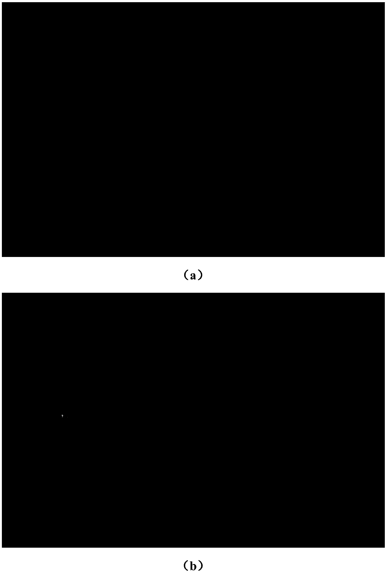
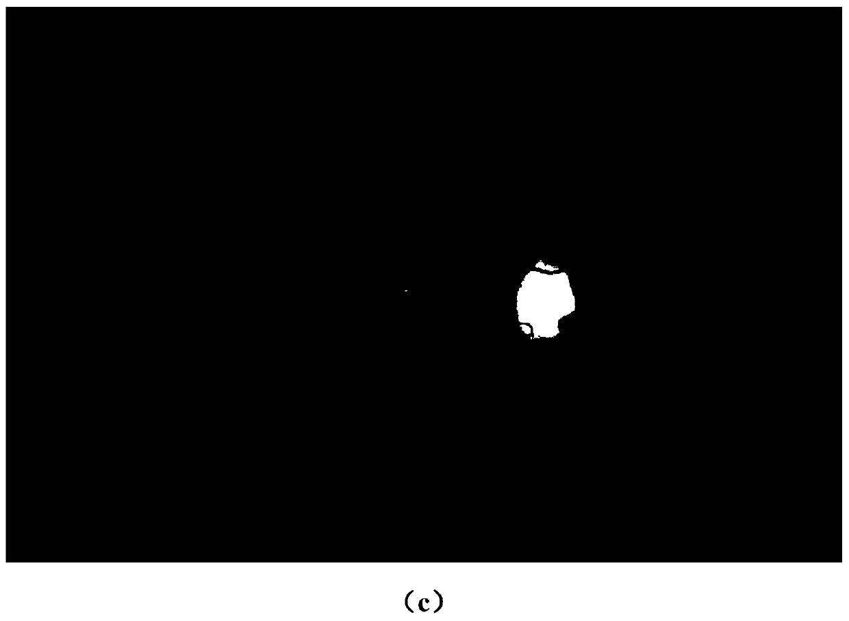
[0030] Embodiment 1, a kind of fundus image optic disc and macular localization detection algorithm based on YOLO-V3, see group figure 1 , 2 , follow the steps below:

[0031] a. Collect and make retinal fundus image data sets with optic disc and macular labels;

[0032] b. After the optic disc and macular data sets of fundus images are produced, modify the network parameters according to the target category to be identified and start the training of the model;

[0033] c. After the model training is completed, multiple sets of independent data set tests are performed to realize rapid fundus image optic disc and macular positioning detection and evaluate the model detection effect.

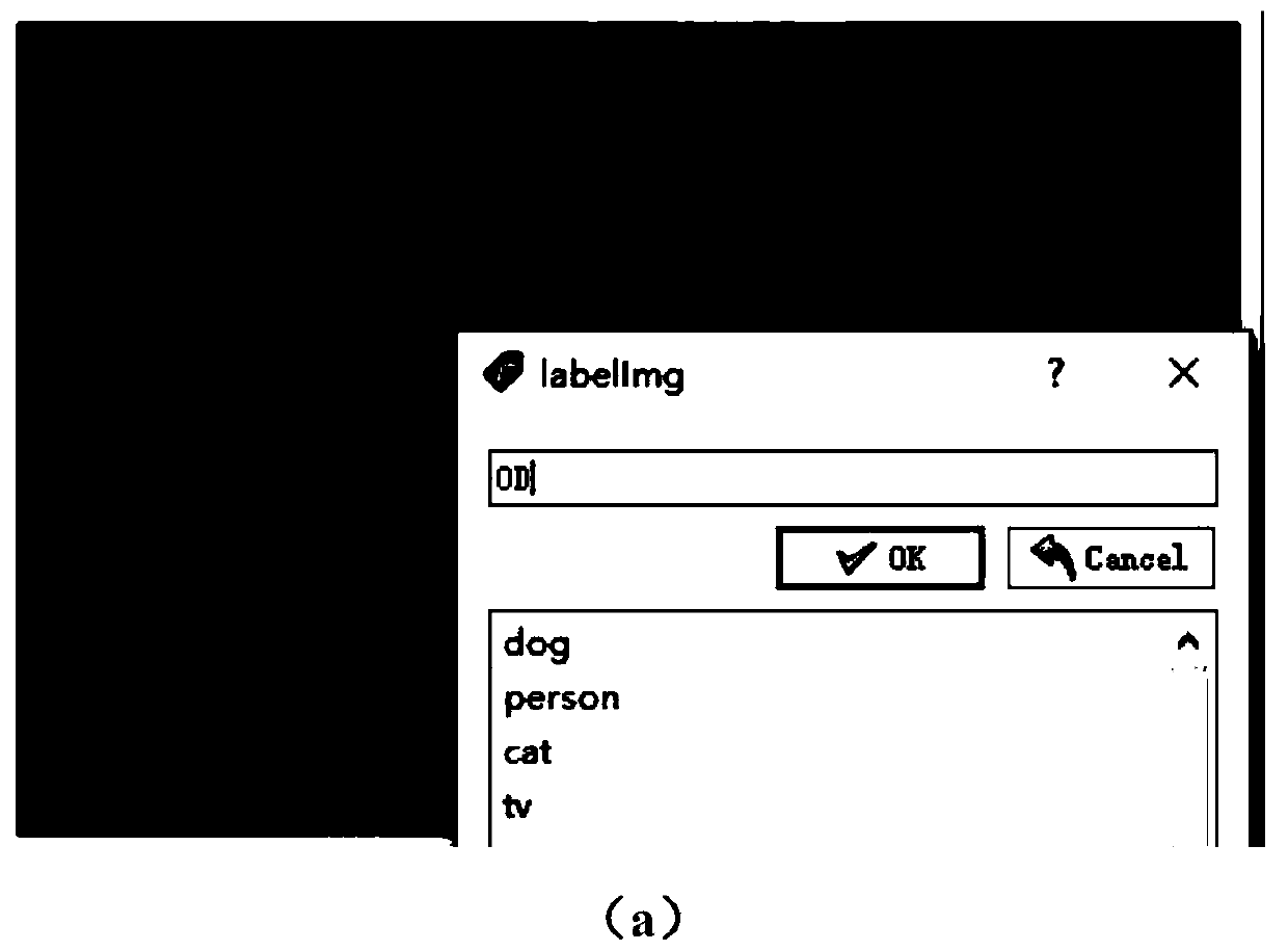
[0034] In the aforementioned YOLO-V3-based fast fundus image optic disc and macular location detection method, in the step a, the format of the retinal fundus image data set is VOC format; the retinal fundus image data set is made according to the following method: identify Collect a certain am...


PUM


Abstract

The invention discloses a n eye fundus image optic disc and macular positioning detection algorithm based on YOLO-V3 in the field of medical image recognition. The eye fundus image optic disc and macular positioning detection algorithm comprises the following steps: a, collecting and manufacturing a retina eye fundus image data set with an optic disc and a macular label; b, after the fundus imageoptic disk and macular data set is manufactured, modifying network parameters according to the target category needing to be recognized, and training a model; and c, after the model training is completed, testing multiple groups of independent data sets to realize rapid fundus image optic disc and macular positioning detection and evaluate a model detection effect. The positions of the optic discand the macula luteae in the fundus image can be simultaneously positioned in one set of system, the identification precision and speed are improved compared with those of a traditional positioning method, and the complexity of a corresponding positioning algorithm is reduced at the same time.

Description

technical field [0001] The invention relates to the technical field of medical image recognition, in particular to a YOLO-V3-based optic disc and macular location detection algorithm for fundus images. Background technique [0002] Fundus retinal examination has been widely used in the prevention, diagnosis and treatment of diabetic retinopathy, glaucoma, age-related macular degeneration and other eye diseases. Fundus images include the optic disc, macula, blood vessels and other major structures, and feature analysis of these structures is the basis for judging fundus diseases. As one of the important structures of the fundus image, the optic disc's size and shape are the main auxiliary parameters for judging various ophthalmic diseases, and are often used as indicators for judging retinopathy. The optic disc is also a key part of detecting other retinal structures. The distance between the optic disc and the macular area is generally fixed, so the center position informa...

Claims


Application Information

Patent Timeline
Patent Type & Authority Applications(China)
IPC IPC(8): G06T7/00
CPCG06T7/0012G06T2207/20084G06T2207/30041
Inventor 张荣芬孙雨琛张达峰刘宇红
Owner GUIZHOU UNIV
Why Eureka
Try Eureka