Target area extraction method and system based on craniocerebral image data

A technology of image data and target area, applied in the fields of medical imaging and computer, to simplify the calculation process and shorten the time-consuming effect

Pending Publication Date: 2020-05-08
UNION STRONG (BEIJING) TECH CO LTD
View PDF5 Cites 9 Cited by
  • Summary
  • Abstract
  • Description
  • Claims
  • Application Information

AI Technical Summary

Problems solved by technology

[0005] The embodiment of this specification provides a method and system for extracting a target area based on brain image data, which is used to solve the following technical problems: it can eliminate or reduce the participation of human factors, simplify the calculation process, shorten time-consuming, and realize simple and fast Evaluation of core infarcts and / or areas of hypoperfusion

Method used


Image

  • Target area extraction method and system based on craniocerebral image data
  • Target area extraction method and system based on craniocerebral image data
  • Target area extraction method and system based on craniocerebral image data

Examples


Embodiment Construction

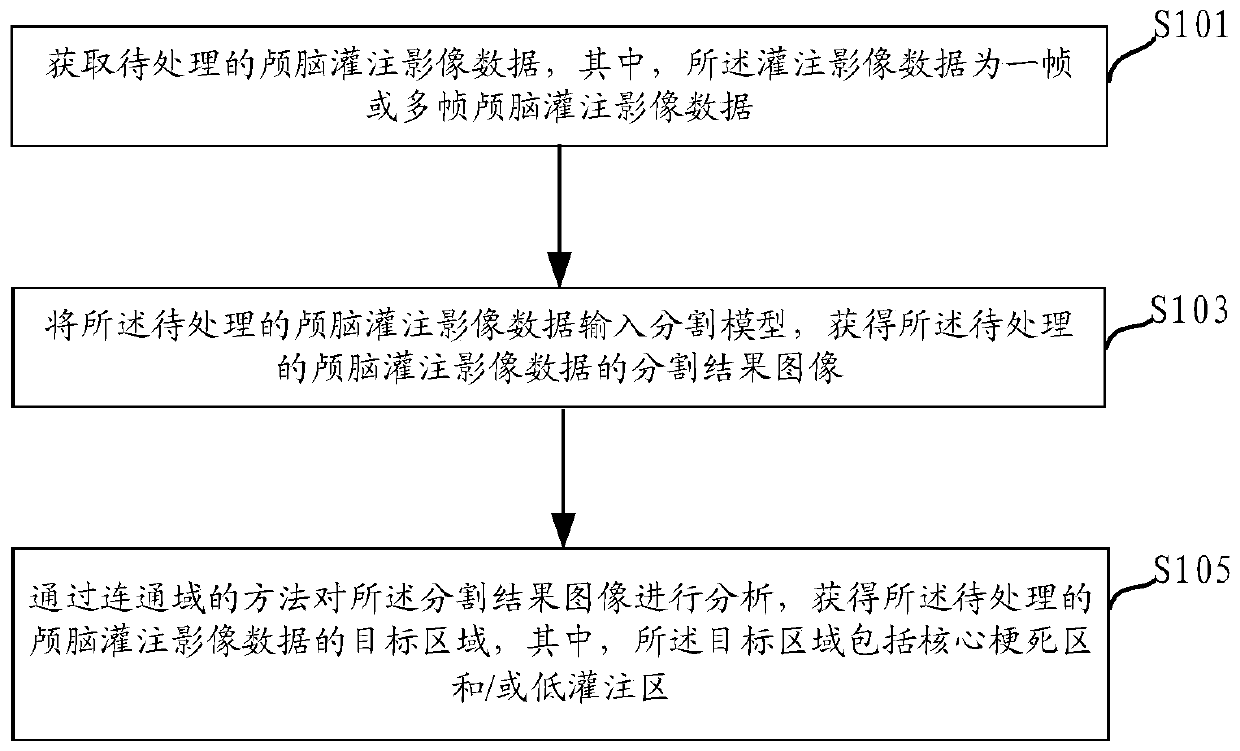
[0032] In order to enable those skilled in the art to better understand the technical solutions in this specification, the technical solutions in the embodiments of this specification will be clearly and completely described below in conjunction with the drawings in the embodiments of this specification. Obviously, the described The embodiments are only some of the embodiments of the present application, but not all of them. Based on the embodiments of this specification, all other embodiments obtained by persons of ordinary skill in the art without creative efforts shall fall within the scope of protection of this application.

[0033] Perfusion imaging using CTP (CT perfusion, CT perfusion imaging) and MRP (MR perfusion, magnetic resonance perfusion imaging) has become a routine method for checking cerebral blood flow in stroke patients. Cerebral perfusion imaging is a continuous dynamic scan of the selected layer of interest to obtain the time density curve of each pixel of...


PUM


Abstract

The embodiment of the invention discloses a target area extraction method and system based on craniocerebral image data, and solves the problems of tedious calculation, long time consumption and the like in the prior art through the extraction method based on the craniocerebral image data. The method comprises the following steps: acquiring to-be-processed craniocerebral perfusion image data; inputting the to-be-processed craniocerebral perfusion image data into a segmentation model to obtain a segmentation result image of the to-be-processed craniocerebral perfusion image data; and analyzingthe segmentation result image through a connected domain method to obtain a target area of the to-be-processed craniocerebral perfusion image data, the target area comprising a core infarction area and / or a hypoperfusion area. The target area can be automatically and directly displayed on the craniocerebral perfusion image, the calculation process is simplified, the consumed time is shortened, andthe evaluation of the core infarction area and / or the hypoperfusion area can be simply and quickly carried out.

Description

technical field [0001] This specification relates to the fields of medical imaging and computer technology, and in particular to a method and system for extracting a target region based on brain imaging data. Background technique [0002] With the development of computer technology, the clinical application of medical imaging has become more and more extensive. Medical imaging provides a great scientific and intuitive basis for the diagnosis of diseases, which can better cooperate with clinical symptoms, laboratory tests, etc., and play an irreplaceable role in the final accurate diagnosis of the disease; at the same time, medical imaging can also be applied In terms of treatment. [0003] The important application of medical imaging technology in brain histology is to screen the indications for endovascular interventional therapy. Firstly, the risk of endovascular interventional therapy is assessed by the extent of the core infarct area, and secondly, by the extent of the ...

Claims


Application Information

Patent Timeline
Patent Type & Authority Applications(China)
IPC IPC(8): G06T7/00G06T7/10
CPCG06T7/0012G06T7/10G06T2207/10081G06T2207/10088G06T2207/30016G06T2207/30104G06T2207/20081G06T2207/20084G06T2207/20132
Inventor 杨鹏飞金海岚刘建民印胤洪波杨光明张永巍秦岚张磊张洪剑张永鑫
Owner UNION STRONG (BEIJING) TECH CO LTD
Why Eureka
Try Eureka