Digital pathological section mitotic image statistical method, device, equipment and medium

A technology of digital pathological slices and silk nucleus, applied in image data processing, calculation, image analysis and other directions, can solve problems such as difference in results

Pending Publication Date: 2020-05-15
GUANGZHOU KINGMED DIAGNOSTICS CENT
View PDF5 Cites 4 Cited by
  • Summary
  • Abstract
  • Description
  • Claims
  • Application Information

AI Technical Summary

Problems solved by technology

However, under the conditions of artificial intelligence reading, statistical methods for mitotic figures may cause differences in results due to factors such as microscope specifications, tumor-to-stroma ratio, and cell density.

Method used


Image

  • Digital pathological section mitotic image statistical method, device, equipment and medium
  • Digital pathological section mitotic image statistical method, device, equipment and medium
  • Digital pathological section mitotic image statistical method, device, equipment and medium

Examples


Embodiment Construction

[0065] The following will clearly and completely describe the technical solutions in the embodiments of the present invention with reference to the accompanying drawings in the embodiments of the present invention. Obviously, the described embodiments are only some, not all, embodiments of the present invention. Based on the embodiments of the present invention, all other embodiments obtained by persons of ordinary skill in the art without creative efforts fall within the protection scope of the present invention.

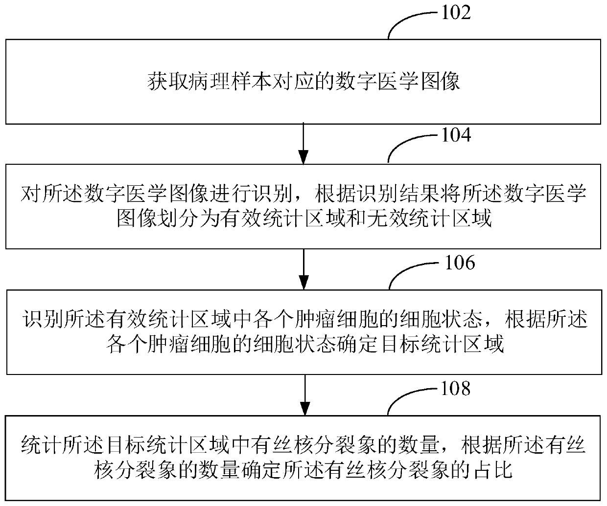
[0066] In one embodiment, such as figure 1 As shown, a method for counting mitotic figures in digital pathological sections is provided, which specifically includes the following steps:

[0067] Step 102, acquiring digital medical images corresponding to pathological samples.

[0068] Among them, the digital medical image refers to the medical image obtained by scanning and in which tumor cells can be directly observed. In order to obtain digital medical images, ...


PUM


Abstract

The embodiment of the invention discloses a digital pathological section mitotic image statistical method. The method comprises the following steps: acquiring a digital medical image corresponding toa pathological sample; identifying the digital medical image, and dividing the digital medical image into an effective statistical area and an invalid statistical area according to an identification result; identifying the cell state of each tumor cell in the effective statistical area, and determining a target statistical area according to the cell state of each tumor cell; and counting the number of the filament nuclear split images in the target statistical area, and determining the proportion of the filament nuclear split images according to the number of the filament nuclear split images.According to the digital pathological section mitotic image statistical method, the number of mitotic images in the statistical target statistical area is determined; therefore, diagnostic junction deviation caused by factors such as microscope specification, tumor stromal ratio and cell density is eliminated, and meanwhile, due to the combination of computer recognition and machine vision, rapidand accurate quantitative detection and analysis can be realized.

Description

technical field [0001] The invention relates to the technical field of mitotic mitosis statistics, in particular to a method, device, equipment and medium for mitotic mitotic statistics of digital pathological slices. Background technique [0002] One of the characteristics of malignant tumors is the increase in the number of mitotic figures in tumor cells, so counting the number of mitotic figures can evaluate the malignancy of the tumor to a certain extent. The usual statistical method of mitotic figures is to find the active areas of mitotic figures, and then start to observe and count the number of mitotic figures in multiple groups of 10-40 high-power fields of view, and finally take the average value of mitotic figures as Final statistics. With the continuous advancement of computer technology, the combination of artificial intelligence and medical treatment is gradually being applied. By combining technologies such as big data and deep learning, artificial intellige...

Claims


Application Information

Patent Timeline
Patent Type & Authority Applications(China)
IPC IPC(8): G06T7/00
CPCG06T7/0012G06T2207/30096G06T2207/10061
Inventor 车拴龙李晶李学锋刘栋吴涛罗丕福刘斯李映华
Owner GUANGZHOU KINGMED DIAGNOSTICS CENT
Why Eureka
Try Eureka