
Display of a medical image

A technology of medical images and image data, applied in the field of medical images, can solve problems such as invisibility, and achieve the effect of easy identification and evaluation, and reduced volume

Pending Publication Date: 2020-05-19
KONINKLJIJKE PHILIPS NV
View PDF4 Cites 0 Cited by
  • Summary
  • Abstract
  • Description
  • Claims
  • Application Information

AI Technical Summary

Problems solved by technology

[0006] Furthermore, even for CT images where standard transfer functions are widely used, the choice of a specific specific transfer function may result in specific abnormalities not being visible, thereby preventing the discovery of possible incidental findings (e.g., liver tumors may be viewed or visualized using dedicated lung tissue). parameter is not visible)

Method used


Image

  • Display of a medical image
  • Display of a medical image
  • Display of a medical image

Examples


Embodiment Construction

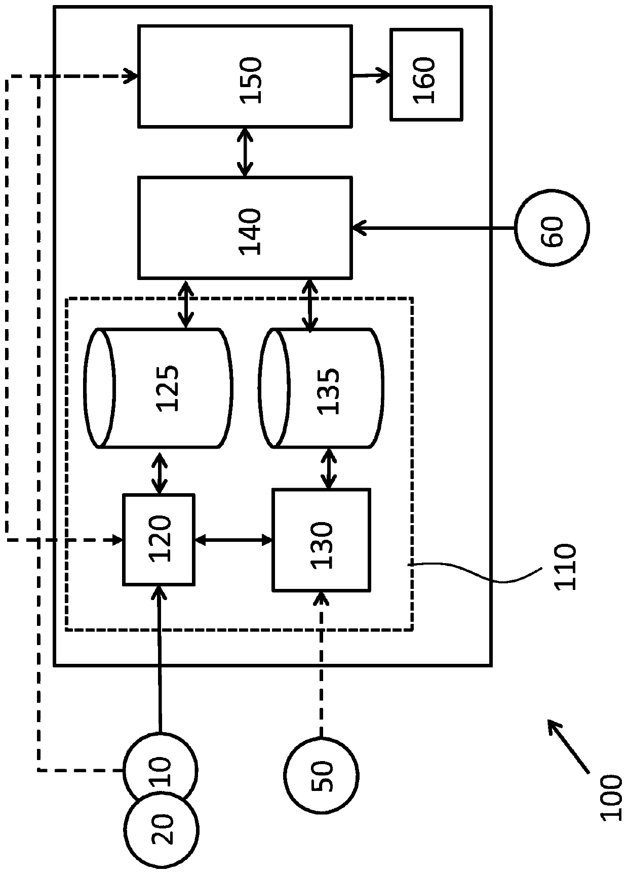
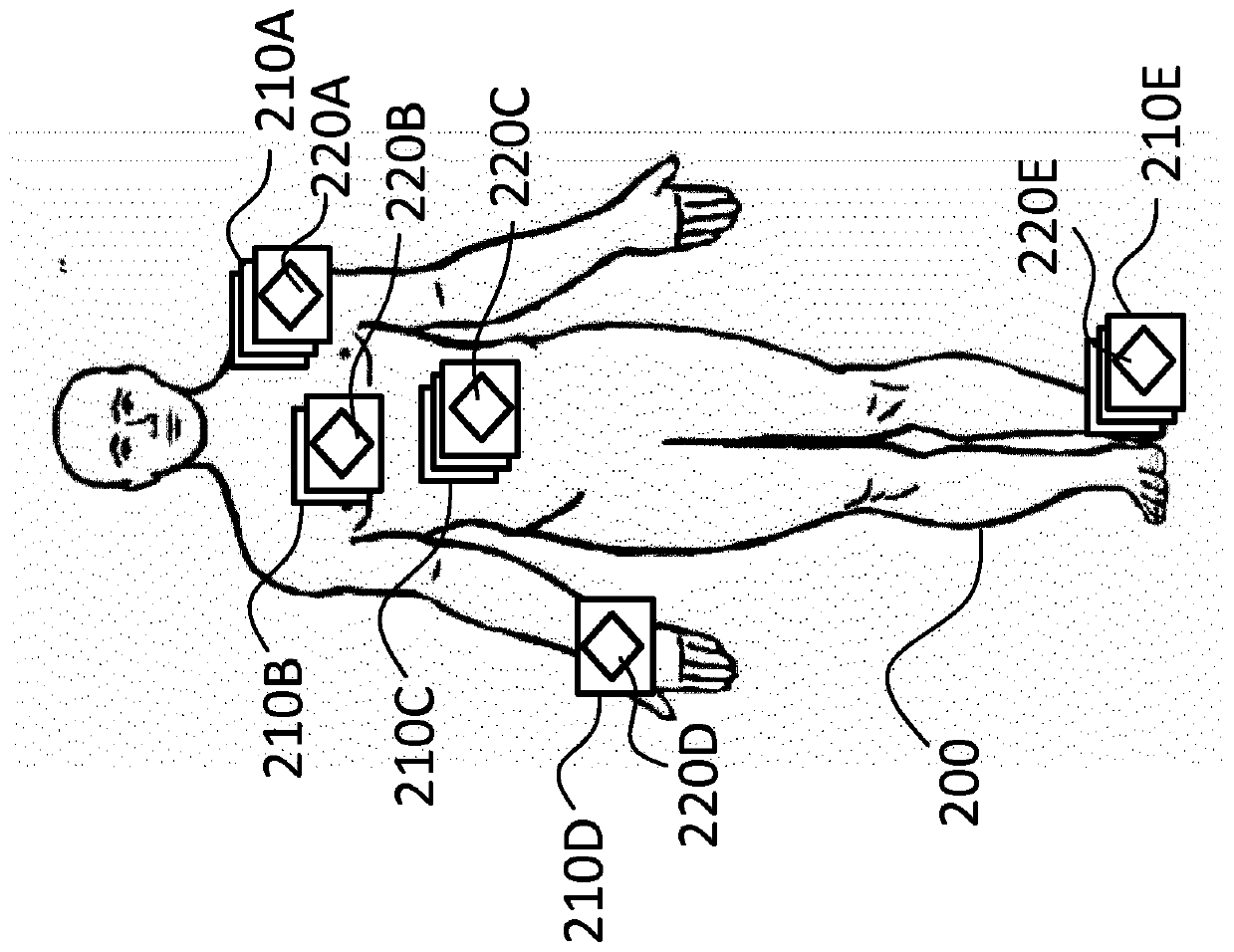
[0045] The proposed embodiments relate to generating transformed data for displaying medical images using optimal or preferred visualization parameters / settings. Medical images can be mapped to an anatomical atlas (e.g., a representation of the relative positions of anatomical features of a human or animal body), and transfer functions (used to transform image data for display according to optimal or preferred visualization parameters) can be associated with the anatomical The location in the atlas is associated. In this way, an anatomical atlas can be supplemented with information for transforming medical image data for display, where the information depends on the position within the atlas reference space.

[0046] Accordingly, some embodiments of the invention may be used in conjunction with medical images obtained using different methods and / or data capture settings. Embodiments may enable automatic selection of transfer functions (for transforming medical image data for ...


PUM


Abstract

Presented are concepts for displaying medical images. One such concept generates transformation data by mapping image data of a medical image to a location in an anatomical atlas and by determining atransfer function for transforming image data of the medical image for display. The transfer function is then associated with the location in the anatomical atlas. A medical image can then be displayed by identifying a transfer function associated with a selected location in the anatomical atlas. The identified transfer function is applied to image data of a medical image that is mapped to the selected location in the anatomical atlas so as to obtain transformed image data. A transformed version of the medical image is then displayed based on the transformed image data.

Description

technical field [0001] The present invention relates to medical images, and more particularly to displaying medical images. Background technique [0002] The increasing number of medical image acquisition techniques inevitably leads to an increasing number of visualization methods and demands to be fulfilled. However, important considerations for viewing and interpreting medical images include reproducibility and consistency across a variety of medical images. Such considerations may be complex and / or intractable, for example when various medical images have been acquired using different techniques, devices, and / or data capture parameters. [0003] For example, to view and assess lung disease in computed tomography (CT) images, the image data can be displayed using specific viewing or visualization parameters that convert the Hounsfield unit (HU) values ​​of the CT image data into gray value of . However, viewing and evaluating skeletal tissue may use other viewing or vis...

Claims


Application Information

Patent Timeline
Patent Type & Authority Applications(China)
IPC IPC(8): G06T1/00
CPCG06T1/00G06T5/009G06T5/50G06T2207/10072G06T2207/30004G06T2207/20128G06T7/73G06T7/0012G06T2200/24G06F18/213
Inventor D·贝斯特罗夫T·维克A·R·弗兰茨H·舒尔茨N·沙德瓦尔特
Owner KONINKLJIJKE PHILIPS NV
Learn More