Application of deep learning for medical imaging evaluation

A deep learning and medical imaging technology, applied in the field of deep learning algorithm development, can solve problems such as algorithm robustness concerns

Pending Publication Date: 2020-07-21
库雷人工智能科技私人有限公司
View PDF2 Cites 0 Cited by
  • Summary
  • Abstract
  • Description
  • Claims
  • Application Information

AI Technical Summary

Problems solved by technology

The training and validation datasets for most studies had <200 head CT scans, raising concerns about the robustness of these algorithms
Furthermore, there is no standard public head CT dataset to directly compare the performance of algorithms

Method used


Image

  • Application of deep learning for medical imaging evaluation
  • Application of deep learning for medical imaging evaluation
  • Application of deep learning for medical imaging evaluation

Examples


example 1

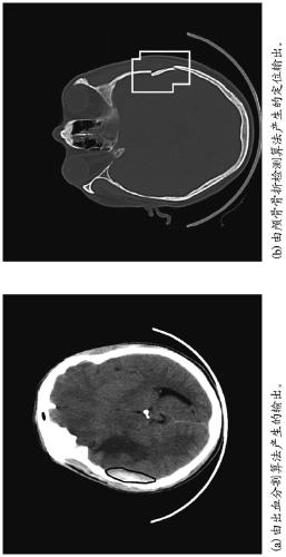
[0052] Example 1. Deep Learning Algorithm for Detecting Key Findings in Head CT Scans

[0053] 1.1 Dataset

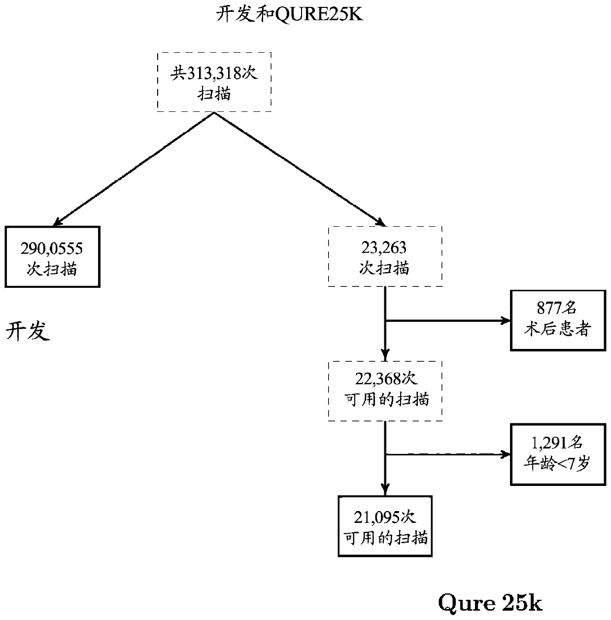
[0054] 313,318 anonymized head CT scans were collected retrospectively from several centers in India. These centers included inpatient and outpatient radiology centers employing a variety of CT scanner models (Table 1), where the number of slices per rotation ranged from 2 to 128. Every scan has an electronic clinical report associated with it, which we used as the gold standard during the algorithm development process.

[0055] Table 1. Models of CT scanners used for each dataset.

[0056]

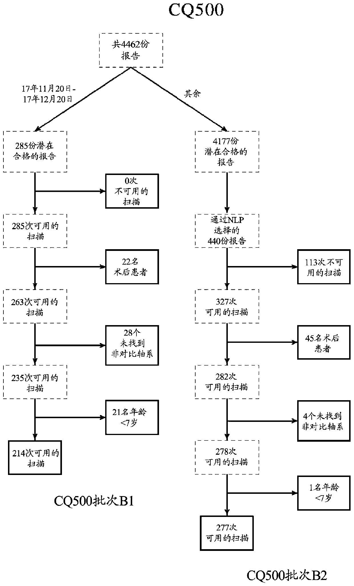
[0057] Of these scans, scans of 23,263 randomly selected patients (Qure25k dataset) were selected for validation, and scans of the remaining patients (development dataset) were used for training / development of the algorithm. Post-operative scans and scans of patients younger than 7 years old were removed from the Qure25k dataset. This dataset was not used during the algorith...


PUM


Abstract

This disclosure generally pertains to methods and systems for processing electronic data obtained from imaging or other diagnostic and evaluative medical procedures. Certain embodiments relate to methods for the development of deep learning algorithms that perform machine recognition of specific features and conditions in imaging and other medical data. Another embodiment provides systems configured to detect and localize medical abnormalities on medical imaging scans by a deep learning algorithm.

Description

[0001] related application [0002] This application claims the benefit of priority from Indian Patent Application No. 201821042894 filed on November 14, 2018, which is hereby incorporated by reference in its entirety for all purposes. technical field [0003] The present disclosure generally relates to methods and systems for processing electronic data obtained from imaging or other diagnostic and evaluation medical procedures. Some embodiments relate to methods for the development of deep learning algorithms that perform machine recognition of specific features and conditions in imaging and other medical data. Background technique [0004] Medical imaging techniques such as computed tomography (CT) and X-ray imaging are widely used in diagnosis, clinical research and treatment planning. There is an emerging need for automated methods that improve the efficiency, accuracy, and cost-effectiveness of medical imaging assessments. [0005] Non-contrast head CT scans are among...

Claims


Application Information

Patent Timeline
Patent Type & Authority Applications(China)
IPC IPC(8): G06T7/00G06K9/62
CPCG06V2201/03G06V10/82G06V10/764
Inventor 萨桑克·奇拉姆库尔希罗希特·高希斯威萨·塔纳马拉普贾·拉奥普拉桑特·瓦瑞尔
Owner 库雷人工智能科技私人有限公司
Why Eureka
Try Eureka