Craniocerebral operation channel

A technology of cranial surgery and access, which is applied in the field of cranial surgery access, can solve the problems of large brain tissue damage and difficulty in locating lesions, and achieve the effect of small trauma and convenient intervention

Pending Publication Date: 2020-07-24
CHONGQING JINYUANXI MEDICAL TECH CO LTD
View PDF0 Cites 1 Cited by
  • Summary
  • Abstract
  • Description
  • Claims
  • Application Information

AI Technical Summary

Problems solved by technology

The existing surgical access is often performed by incision and ostomy + brain compression plate traction, which causes great damage to the brain tissue
In addition, it is difficult to locate the lesion hidden in the brain parenchyma (like digging a well in a field)

Method used


Image

  • Craniocerebral operation channel
  • Craniocerebral operation channel
  • Craniocerebral operation channel

Examples


Embodiment 1

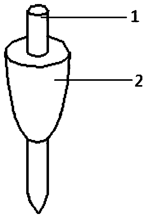
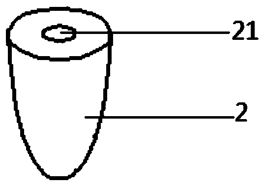
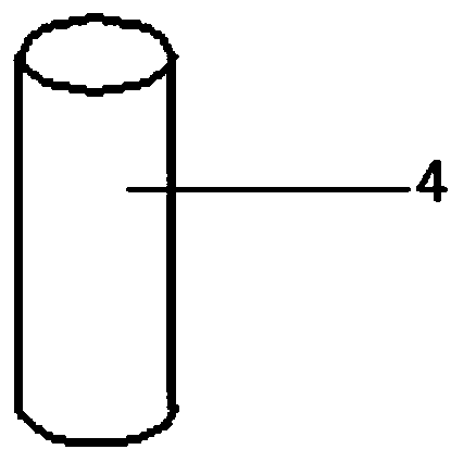
[0027] Such as Figure 1-3 As shown, the present embodiment provides a craniocerebral operation channel, including a guide device, a puncture cone 2 and an outer sheath tube 4, the guide device is used to penetrate into the patient's brain, and penetrates into the patient's brain before the operation channel is established, and After reaching the lesion position, the puncture can play a guiding role in the establishment of the subsequent surgical channel. The middle part of the puncture cone 2 is provided with a communication hole 21, and the communication hole 21 runs through the puncture cone 2 in the direction of the central axis of the puncture cone 2, and the communication hole 21 Set coaxially with the puncture cone 2, the puncture cone 2 can be sleeved on the outer periphery of the guide device through the communication hole 21, and the puncture cone 2 can slide in the length direction of the guide device, when the guide device punctures in place, slowly insert the punct...

Embodiment 2

[0034] Such as Figure 4-6 As shown, the difference between this embodiment and Embodiment 1 is that the guiding device is a neuronavigation probe 3, and neuronavigation is a precise positioning technology for preoperatively designing a surgical plan and guiding a surgical operation in real time during the operation. Realize precise positioning.

[0035] The puncture cone 2 is also provided with a communication groove 22, the communication groove 22 runs through the puncture cone 2 in the central axis direction of the puncture cone 2, and the groove bottom of the communication groove 22 extends to the inner wall of the communication hole 21, and the opening of the communication groove 22 extends to The side wall of the puncture cone 2 can be sleeved on the outer periphery of the neuronavigation probe 3 through the opening of the communication groove 22. Since there are other devices on the end of the neuronavigation probe 3 away from the needle tip, it is impossible to pass th...


PUM


Abstract

The invention discloses a craniocerebral operation channel, belonging to the technical field of medical instruments. The craniocerebral operation channel comprises a guiding device, a trocar and an outer sheath tube, wherein the guiding device is used for penetrating into the brain of a patient; a communicating hole is formed in the middle part of the trocar; the communicating hole penetrates through the trocar in the direction of the central axis of the trocar and is coaxially arranged with the trocar; the trocar can be arranged on the periphery of the guiding device in a sleeving mode through the communicating hole and can slide in the length direction of the guiding device; the interior of the outer sheath tube is hollow and used for providing an operation channel; and the outer sheathtube can be arranged on the periphery of the trocar in a sleeving mode. The craniocerebral operation channel is accurate in positioning and can effectively protect the brain tissue of the patient.

Description

technical field [0001] The invention relates to the technical field of medical instruments, in particular to a cranial operation channel. Background technique [0002] Craniocerebral surgery under neuroendoscopy has become an important direction in the development of minimally invasive neurosurgery. The core of this technology is to further reduce the trauma of previous surgical approaches, expand the exposure of the disease, and increase the chance of visually resecting the lesion. Since the brain is a solid spherical tissue, in order to reach the lesion in the brain parenchyma during surgery, it is necessary to artificially establish a surgical channel. The existing surgical access is often performed by incision and ostomy + brain compression plate traction, which causes great damage to the brain tissue. In addition, it is difficult to locate the lesion hidden in the brain parenchyma (like digging a well in a field). Contents of the invention [0003] The purpose of th...

Claims


Application Information

Patent Timeline
Patent Type & Authority Applications(China)
IPC IPC(8): A61B17/34
CPCA61B17/3403A61B17/3472A61B17/3421A61B17/00234A61B17/3417A61B2090/103A61B17/3496A61B2017/3456A61B2017/3405
Inventor 李瑛
Owner CHONGQING JINYUANXI MEDICAL TECH CO LTD
Why Eureka
Try Eureka